Article
Why Bulldogs Get Cherry Eye More Often: What Every Veterinarian Should Know About Genetics
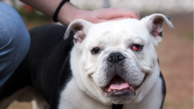
Every small animal practitioner has noticed the pattern. A young English Bulldog presents with a pink mass protruding from the medial canthus. A few weeks later, another Bulldog—or perhaps a French Bulldog or Cocker Spaniel—arrives with the same condition. While cherry eye has traditionally been attributed to weak connective tissue supporting the third eyelid gland, recent genetic discoveries suggest the explanation runs much deeper.
A landmark genome-wide association study published in 2024 identified the FGF4L1 retrogene insertion as a significant genetic risk factor for prolapse of the gland of the nictitating membrane, providing the strongest evidence to date that cherry eye has a hereditary basis1. For veterinarians, this finding has important implications for diagnosis, owner counselling, and breeding recommendations.
Beyond anatomy: understanding the genetic link
For decades, cherry eye was believed to result primarily from laxity or weakness of the connective tissue anchoring the third eyelid gland. Although this mechanical explanation remains valid, genetics now appears to be one of the underlying drivers of that structural weakness.
The 2024 study demonstrated that dogs carrying the FGF4L1 retrogene insertion had significantly higher odds of developing cherry eye than unaffected dogs. Researchers proposed that the mutation may influence connective tissue integrity, making the gland more susceptible to prolapse even under normal physiological forces1.
Earlier pedigree investigations had already shown that the disease follows a complex pattern of inheritance, rather than simple dominant or recessive transmission. This means multiple genes, together with breed-specific anatomy and environmental influences, likely contribute to disease development2.
For clinicians, the message is clear: cherry eye is not simply an isolated ocular disorder—it is a genetically influenced condition.
Which breeds deserve closer attention?
Although cherry eye can occur in any dog, certain breeds consistently show a much higher prevalence.
Commonly affected breeds include:
- English Bulldog
- French Bulldog
- American Cocker Spaniel
- Beagle
- Lhasa Apso
- Shih Tzu
- Cane Corso
- Neapolitan Mastiff
Among these, Bulldogs are frequently overrepresented in clinical studies because of their inherited craniofacial conformation and connective tissue characteristics1,2.
Veterinarians should recognise that young dogs from predisposed breeds presenting for routine health checks may already have mild gland instability before obvious prolapse develops.
Genetics should influence clinical conversations
Understanding the hereditary nature of cherry eye changes how veterinarians communicate with owners.
When discussing treatment, explain that surgery corrects the clinical manifestation of the disease but does not eliminate the underlying genetic predisposition. Consequently, even after successful gland repositioning, some dogs remain at risk of recurrence or later prolapse of the opposite eye1,2.
Owners should therefore be advised to:
- Monitor the contralateral eye regularly.
- Seek veterinary attention promptly if swelling develops.
- Attend recommended postoperative rechecks.
- Understand that recurrence is not always the result of surgical failure.
This discussion helps establish realistic expectations and improves compliance with long-term follow-up.
Should affected dogs be bred?
The growing body of genetic evidence has important implications for breeding programmes.
Although no routine genetic screening test is currently available for cherry eye, researchers suggest that breeders should exercise caution when selecting breeding animals from affected bloodlines. Since pedigree studies indicate a hereditary component and the FGF4L1 retrogene has now been identified as a significant risk factor, reducing the frequency of affected animals in breeding populations may help lower disease prevalence over time1,2.
Veterinarians play an important advisory role by encouraging responsible breeding practices while explaining that cherry eye is likely influenced by multiple genes rather than a single mutation.
Genetics does not replace good surgery
While genetics influences which dogs develop cherry eye, it does not diminish the importance of appropriate surgical management.
Early gland-preserving procedures, particularly Morgan's Pocket Technique, remain the treatment of choice because they preserve tear production and reduce the likelihood of keratoconjunctivitis sicca3. However, clinicians should remember that recurrence in genetically predisposed dogs may reflect underlying connective tissue biology rather than deficiencies in surgical technique.
Combining evidence-based surgery with informed owner counselling provides the best opportunity for long-term success.
Clinical Pearl
Every case of cherry eye has two components: a prolapsed gland that can be repaired surgically and a genetic predisposition that cannot. Understanding both allows veterinarians to provide better treatment, realistic prognostic advice, and responsible breeding recommendations.
References (Vancouver)
- Freyer J, Labadie JD, Huff JT, et al. Association of FGF4L1 Retrogene Insertion with Prolapsed Gland of the Nictitans (Cherry Eye) in Dogs. Genes. 2024;15(2):198. Available from: https://pmc.ncbi.nlm.nih.gov/articles/PMC10887708/
- O'Neill DG, Church DB, McGreevy PD, et al. Investigating the inheritance of prolapsed nictitating membrane glands in a large canine pedigree. Canine Genetics and Epidemiology. 2015;2:7. Available from: https://pmc.ncbi.nlm.nih.gov/articles/PMC4361898/
- White C, Brennan ML. An evidence-based rapid review of surgical techniques for correction of prolapsed nictitans glands in dogs. Vet Sci. 2018;5(3):75. Available from: https://pmc.ncbi.nlm.nih.gov/articles/PMC6163435/
Related Contents

Upcoming Event
African Swine Fever: Diagnosis and Management
African swine fever can cause severe disease and significant losses in affected pig populations.&nbs...
.jpg)
Video
Transjugular Removal of Canine Heartworms: A Clinical and Surgical Overview
Transjugular removal of canine heartworms is a specialized intervention used in selected dogs with s...

Article
Feline Pregnancy Endocrinology: Hormonal Changes and Placental Function
Pregnancy in cats involves a distinctive endocrine pattern in which ovarian and placental hormones w...

Article
Hormonal Control of Pregnancy Termination in Cats
Pregnancy maintenance in cats depends strongly on coordinated hormonal support, particularly progest...

Article
Feline Embryo Implantation and Early Placental Development
Pregnancy establishment in cats involves a sequence of precisely timed events between fertilization,...

Article
Ultrasonographic Monitoring of Pregnancy in Cats: A Gestational Timeline
Ultrasonography provides a practical way to follow the progression of feline pregnancy by identifyin...

Article
Feline Mismating: Pregnancy Diagnosis and Clinical Management
Mismating or unplanned mating is a common clinical problem in sexually mature queen cats because of...

Article
Medical Termination of Pregnancy in Queens: Drugs, Timing and Treatment Protocols
Medical termination of pregnancy (MTP) in queen cats should be approached according to the timing of...