Article
Otoscopy Done Right: Common Findings You Should Never Miss
Is your otoscope helping you diagnose the disease—or simply confirming that the ear is inflamed?
Head shaking, scratching and ear discharge may tell you that a dog has otitis, but they rarely explain why.
That's where otoscopy becomes indispensable.
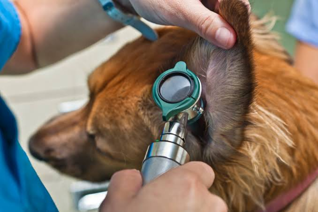
A systematic otoscopic examination not only confirms the presence of otitis externa but also identifies anatomical changes, foreign bodies, masses and tympanic membrane abnormalities that influence both diagnosis and treatment. Performed correctly, otoscopy transforms a routine ear examination into one of the most valuable diagnostic procedures in small animal practice1,2,3.
Otoscopy Should Be More Than a Quick Look
One of the most common mistakes in practice is using the otoscope merely to confirm inflammation.
Instead, every examination should answer five clinical questions:
- Is the ear canal open or narrowed?
- What type of discharge is present?
- Is there evidence of chronic structural change?
- Is a foreign body or mass present?
- Can the tympanic membrane be visualised?
Following the same sequence during every examination reduces the likelihood of missing clinically important findings.
Start with the Pinna
The examination begins before the otoscope enters the ear canal.
Inspect both pinnae for evidence of dermatological disease, including:
- Erythema
- Alopecia
- Excoriations
- Crusting
- Hyperpigmentation
- Swelling
- Pain on palpation
Lesions affecting the pinna often provide valuable clues to underlying diseases such as atopic dermatitis, food allergy or ectoparasitic infestations1.
Assess the Vertical and Horizontal Ear Canal
Once the otoscope is introduced, carefully evaluate the entire ear canal.
Look for:
- Diffuse erythema
- Oedema
- Excess cerumen
- Purulent discharge
- Ulceration
- Haemorrhage
- Foreign material
The appearance of the discharge may also provide useful clinical clues.
- Dry, dark brown wax commonly accompanies ceruminous otitis.
- Thick yellow or green purulent exudate is more often associated with bacterial infection.
- Greasy brown discharge frequently accompanies Malassezia overgrowth.
Although discharge characteristics can support clinical suspicion, they should never replace cytological evaluation1.
Evaluate Canal Diameter
The width of the ear canal provides important information regarding disease chronicity.
Mild acute otitis usually presents with minimal narrowing.
In contrast, recurrent inflammation may result in:
- Oedema
- Glandular hyperplasia
- Fibrosis
- Canal stenosis
Marked narrowing not only makes examination difficult but may also reduce the effectiveness of topical medications by limiting their penetration into the horizontal canal1,3.
Progressive stenosis should alert clinicians to investigate underlying primary disease and consider referral if medical management becomes ineffective.
Never Ignore a Foreign Body
Grass awns remain one of the most common foreign bodies identified during otoscopic examination.
Foreign bodies should be suspected when dogs present with:
- Sudden onset head shaking
- Acute unilateral otitis
- Severe pain
- Minimal previous history of ear disease
Failure to identify and remove the foreign body often results in persistent inflammation despite appropriate antimicrobial therapy.
Look Carefully for Masses
Not every narrowed ear canal is caused by inflammation.
Polyps, ceruminous gland adenomas, neoplasia and other proliferative lesions may partially or completely obstruct the ear canal.
These patients frequently present with:
- Recurrent unilateral otitis
- Persistent discharge
- Poor response to treatment
- Progressive canal obstruction
Any unexplained mass should be investigated further through advanced imaging, biopsy or referral where appropriate.
Can You See the Tympanic Membrane?
Assessment of the tympanic membrane is one of the most important objectives of otoscopy.
Whenever visible, evaluate:
- Integrity
- Colour
- Position
- Opacity
An intact tympanic membrane reduces the likelihood of middle ear involvement but does not completely exclude otitis media1.
Conversely, a ruptured or absent tympanic membrane should prompt careful consideration of:
- Otitis media
- Previous trauma
- Chronic infection
- Selection of non-ototoxic topical medications
If excessive debris prevents adequate visualisation, cleaning and re-examination at a later visit may provide a safer assessment than attempting forceful instrumentation.
When Otoscopy Becomes Difficult
Not every ear can be adequately examined during the first consultation.
Severe pain, marked stenosis, heavy debris or patient anxiety may limit visualisation.
In these situations, clinicians should avoid aggressive manipulation.
Instead, consider:
- Appropriate analgesia
- Sedation or general anaesthesia
- Ear flushing under controlled conditions
- Video otoscopy for complex or chronic cases
A complete examination under sedation often provides substantially more diagnostic information than a painful, incomplete examination in the consulting room.
Otoscopy and Cytology Work Best Together
Otoscopy identifies structural abnormalities.
Cytology identifies microbial populations.
Neither replaces the other.
For example:
- A normal-appearing canal may still contain significant bacterial or yeast overgrowth.
- Conversely, severe canal inflammation may occur with relatively few microorganisms if allergy is the primary driver.
Combining both examinations provides a far more accurate assessment than relying on either technique alone.
Common Otoscopic Mistakes
Avoid these common errors:
- Examining only one ear.
- Failing to inspect the pinna before inserting the otoscope.
- Assuming discharge appearance identifies the pathogen.
- Forgetting to evaluate the tympanic membrane.
- Missing foreign bodies hidden beneath cerumen.
- Treating chronic stenosis as simple acute otitis.
- Skipping otoscopy because the ear appears too painful.
A systematic examination greatly reduces diagnostic errors.
Clinical Pearl
The otoscope should do far more than confirm that an ear is infected.
It should help answer why the disease developed, how advanced it has become and whether additional diagnostics or referral are required.
Key Takeaways
Otoscopy is an essential component of every canine otitis examination. Evaluate the pinna, ear canal, discharge, canal diameter, foreign bodies, masses and tympanic membrane systematically. Chronic structural changes suggest longstanding disease and may alter treatment decisions. Appearance alone cannot distinguish bacterial from yeast infections—combine otoscopy with ear cytology. Sedation or video otoscopy should be considered when pain or severe stenosis prevents adequate examination.
References
- Bajwa J. Canine otitis externa—Treatment and complications. Canadian Veterinary Journal. 2019;60(1):98–101. PMCID: PMC6294027. Available from: https://pmc.ncbi.nlm.nih.gov/articles/PMC6294027/
- Ponn PC, Tipold A, Volk AV, et al. Can We Minimize the Risk of Dogs Developing Canine Otitis Externa? Animals. 2024;14(17):2537. DOI: 10.3390/ani14172537. PMCID: PMC11394639. Available from: https://pmc.ncbi.nlm.nih.gov/articles/PMC11394639/
- Leonard, C.; Thiry, D.; Taminiau, B.; Daube, G.; Fontaine, J. External Ear Canal Evaluation in Dogs with Chronic Suppurative Otitis Externa: Comparison of Direct Cytology, Bacterial Culture and 16S Amplicon Profiling. Vet. Sci. 2022, 9, 366. https://doi.org/10.3390/vetsci9070366
Related Contents

Upcoming Event
African Swine Fever: Diagnosis and Management
African swine fever can cause severe disease and significant losses in affected pig populations.&nbs...
.jpg)
Video
Transjugular Removal of Canine Heartworms: A Clinical and Surgical Overview
Transjugular removal of canine heartworms is a specialized intervention used in selected dogs with s...

Article
Feline Pregnancy Endocrinology: Hormonal Changes and Placental Function
Pregnancy in cats involves a distinctive endocrine pattern in which ovarian and placental hormones w...

Article
Hormonal Control of Pregnancy Termination in Cats
Pregnancy maintenance in cats depends strongly on coordinated hormonal support, particularly progest...

Article
Feline Embryo Implantation and Early Placental Development
Pregnancy establishment in cats involves a sequence of precisely timed events between fertilization,...

Article
Ultrasonographic Monitoring of Pregnancy in Cats: A Gestational Timeline
Ultrasonography provides a practical way to follow the progression of feline pregnancy by identifyin...

Article
Feline Mismating: Pregnancy Diagnosis and Clinical Management
Mismating or unplanned mating is a common clinical problem in sexually mature queen cats because of...

Article
Medical Termination of Pregnancy in Queens: Drugs, Timing and Treatment Protocols
Medical termination of pregnancy (MTP) in queen cats should be approached according to the timing of...