Article
Iron Deficiency Anemia in Dogs: A Practical Clinical Approach to Diagnosis and Management
Iron deficiency anemia (IDA) is an important differential diagnosis in dogs presenting with chronic weakness, pallor, or unexplained weight loss. Rather than being a primary disease, it often reflects an underlying pathological process that results in chronic blood loss or inadequate iron availability. As a microcytic hypochromic anemia, IDA requires veterinarians to move beyond simply correcting hematological abnormalities and focus on identifying the primary cause. Iron plays a vital role in oxygen transport and cellular metabolism, while its absorption and release into circulation are tightly regulated to maintain systemic iron balance1,2,3.
Clinical Evaluation Begins with Recognizing the Pattern1
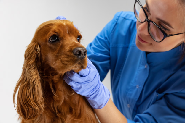
The clinical presentation of IDA is often non-specific, making a thorough physical examination essential. Affected dogs commonly present with lethargy, melena, weight loss, anorexia, inappetence, vomiting, and reduced exercise tolerance. Pica may also be observed in some cases. During physical examination, pale mucous membranes are a prominent finding, frequently accompanied by enlarged lymph nodes, tachycardia, tachypnea, and occasionally hyperthermia. These findings should prompt further investigation to identify the source of chronic iron loss rather than attributing the anemia to nutritional deficiency alone.
Confirming the Diagnosis1
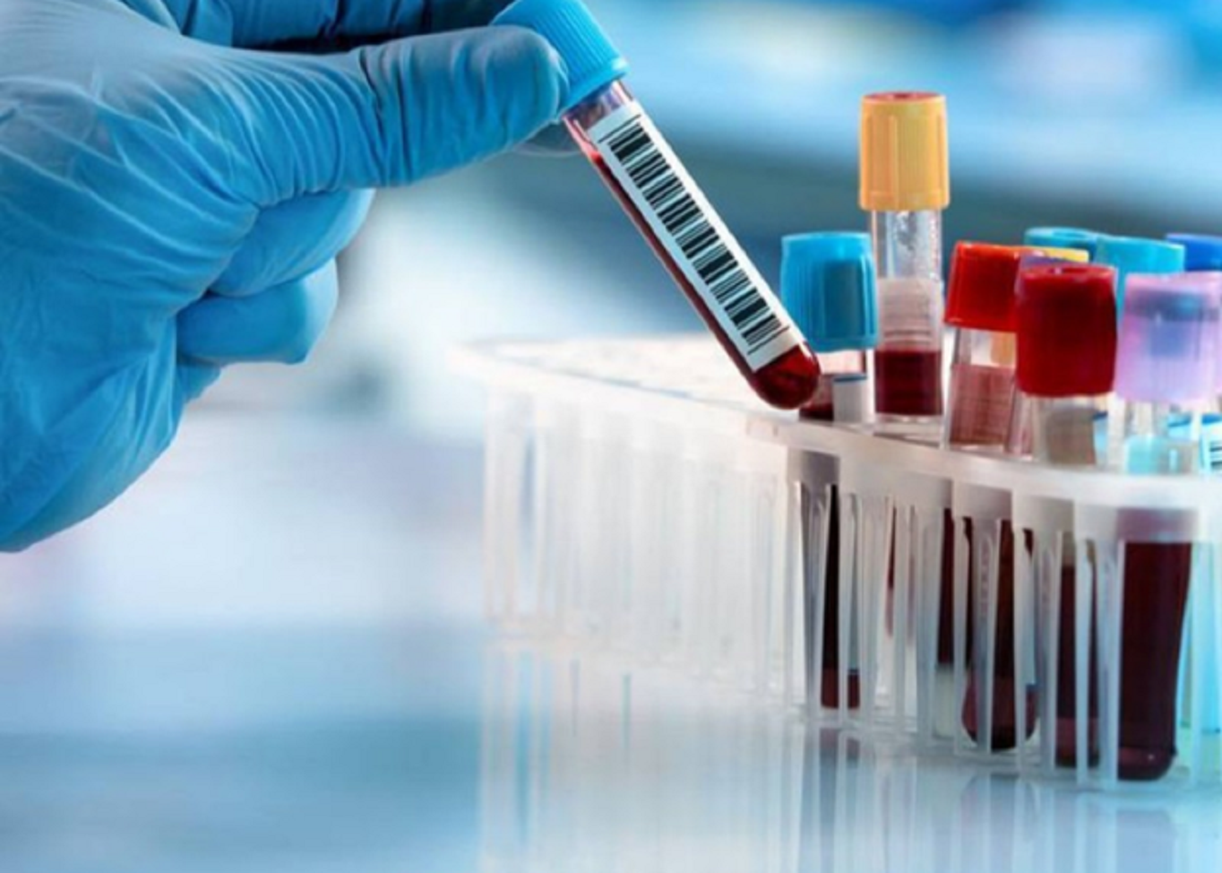
Diagnosis should combine clinical assessment with laboratory evaluation. A complete blood count, peripheral blood smear examination, faecal examination, and serum biochemical analysis provide valuable diagnostic information. Typical laboratory findings include reduced haemoglobin, packed cell volume (PCV), and total erythrocyte count (TEC), consistent with microcytic hypochromic anemia. Evaluation of the iron profile further strengthens the diagnosis by assessing serum iron, total iron-binding capacity (TIBC), unsaturated iron-binding capacity (UIBC), and percentage transferrin saturation (%TSAT), calculated as (Serum iron/TIBC) × 100. Serial assessment of these parameters on follow-up helps monitor therapeutic response and recovery.
Identifying the Underlying Cause is Key1
Successful management depends on addressing the primary condition responsible for iron deficiency. Several underlying etiologies were encountered, including Ancylostoma caninum, gastritis, Babesia gibsoni, demodicosis, haematuria, Dipylidium spp., Toxocara spp., NSAID toxicity, dietary deficiency, and abnormal masses. Therapeutic interventions were directed at these primary disorders using appropriate veterinary protocols. Supportive management included multiple electrolytes, dextrose injections, papaya extract, and liver protectants where required. This comprehensive approach supports hematological recovery while reducing the likelihood of continued iron loss.
Monitoring Response to Therapy1
Treatment protocols included an allopathic regimen consisting of intramuscular iron dextran administered weekly for two weeks, followed by oral ferrous glycinate, copper glycinate, and cobalt glycinate for 21 days. An alternative protocol utilized Ferrum metallicum, Cobaltum metallicum, and Cuprum metallicum over the same treatment period. Clinical progress was evaluated on days 14 and 21 through repeat hematological and biochemical testing.
Both treatment approaches demonstrated progressive improvement in haemoglobin, PCV, TEC, serum iron concentration, and %TSAT, together with reductions in UIBC. The allopathic regimen produced a comparatively faster improvement in haemoglobin, PCV, TEC, and iron parameters, while the alternative regimen also showed consistent improvement throughout the treatment period. Regular laboratory monitoring therefore remains an important component of case management, allowing veterinarians to objectively assess recovery alongside clinical improvement.
Practical Clinical Insights
Iron deficiency anemia should be approached as a clinical indicator rather than a final diagnosis. Dogs presenting with pallor, chronic lethargy, melena, or unexplained weight loss warrant a systematic diagnostic work-up that includes hematological evaluation, iron profile assessment, peripheral blood smear examination, and faecal analysis. Equally important is identifying and treating the underlying cause responsible for chronic iron loss.
Follow-up evaluation using haemoglobin, PCV, TEC, serum iron, UIBC, and %TSAT provides objective evidence of therapeutic response and helps guide ongoing clinical decision-making. By combining targeted treatment of the primary disorder with appropriate correction of iron deficiency, veterinarians can achieve meaningful clinical and hematological improvement while ensuring a more comprehensive approach to patient management.
References
- Jadhav S, Ambore B, Meshram M, Mote C, Jadhav S. Iron deficiency anemia in dogs: Therapeutic management. Int J Adv Biochem Res. 2025;9(Suppl 6):36-40. doi:10.33545/26174693.2025.v9.i6Sa.4489 https://www.doi.org/10.33545/26174693.2025.v9.i6Sa.4489
- Naigamwalla DZ, Webb JA, Giger U. Iron deficiency anemia. The Canadian Veterinary Journal. 2012 Mar;53(3):250. https://pmc.ncbi.nlm.nih.gov/articles/PMC3280776/pdf/cvj_03_250.pdf
- Knovich MA, Storey JA, Coffman LG, Torti SV, Torti FM. Ferritin for the clinician. Blood reviews. 2009 May 1;23(3):95-104. https://pmc.ncbi.nlm.nih.gov/articles/PMC2717717/pdf/nihms113591.pdf
Related Contents

Article
The Elephant is Walking Differently: Where Should the Investigation Begin?
When an elephant begins walking differently, the first challenge is not necessarily to identify the...
Article
When an Elephant’s Blood Report Says “Abnormal”: How Should It Be Interpreted?
A laboratory report can flag a value as abnormal even when an elephant appears clinically healthy. C...
.jpg)
Upcoming Event
World Animal Day
World Animal Day highlights the importance of protecting animal health and welfare through responsib...
Video
Rabies: Epitome of the One Health Paradigm
Rabies exemplifies the One Health approach, linking animal health, human health, veterinary services...
Video
One Health Webinar on Important Updates on Rabies for Veterinary Practitioners and Students
Rabies remains a major zoonotic disease requiring vigilance across veterinary practice and public he...
.jpg)
Video
Transjugular Removal of Canine Heartworms: A Clinical and Surgical Overview
Transjugular removal of canine heartworms is a specialized intervention used in selected dogs with s...

Article
Feline Pregnancy Endocrinology: Hormonal Changes and Placental Function
Pregnancy in cats involves a distinctive endocrine pattern in which ovarian and placental hormones w...

Article
Hormonal Control of Pregnancy Termination in Cats
Pregnancy maintenance in cats depends strongly on coordinated hormonal support, particularly progest...