Article
Diagnosing MUO in Practice: MRI, CSF, and When to Biopsy
No fever, Bloodwork normal, but still, a small-breed dog is acting strangely, presenting with sudden seizures and circling. Now, you are standing at a clinical crossroads: is it Meningoencephalitis of unknown origin (MUO) or an infection you can’t afford to miss?
MUO is one of the most common causes of acute-onset brain disease in small-breed dogs. Ruling out MOU is easy, but confirming it without overlooking a treatable condition is a challenging task.
Let’s explore the stepwise diagnostic approach, which is smart, safe, and effective.
- Start with Suspicion: Identify the Red Flags
MOU can easily be mistaken for a seizure disorder or stroke. Breeds such as Pug, Maltese, and Yorkshire Terriers are mainly affected by MUO1.
Clinical signs reflect focal or multifocal CNS lesions.
- Common neurological signs include2:
- Seizures
- Ataxia
- Proprioceptive deficits
- Vestibular signs
- Blindness
- Multiple cranial nerve deficits
- Head or neck pain (occasionally)
- Systemic signs like fever are rare
- GME typically affects the forebrain, brainstem, or both
- GME can rarely present as meningomyelitis, causing:
- Paresis
- Spinal ataxia
- Spinal cord-only involvement in ~9.5%–13% of cases
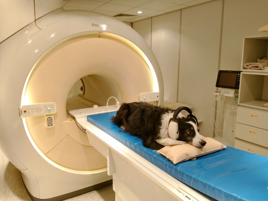
- MRI: The First Line of Insight
MRI is the most crucial step in diagnosing MOU. MRI findings include asymmetrical, multifocal T2 hyperintensities located in the forebrain or cerebellum 3.
It’s important to look for2
- GME: Periventricular or cerebellar white matter lesions
- NME: Gray matter necrosis (Pugs, Maltese)
- NLE: Bilateral white matter damage (Yorkies)
Pro Tip: Always include contrast-enhanced T1-weighted sequences in the MRI protocol to visualize a mild, focal, unilateral temporal muscle lesion 4.
Mistake to Avoid: Don’t rely solely on MRI for diagnosis—MUO can mimic other CNS diseases radiographically5
- CSF Analysis: Timing is everything
Cerebrospinal Fluid (CSF) helps to identify inflammatory changes and rule out infections. CSF key findings include elevated protein and predominantly mononuclear pleocytosis 2.
Key points to keep in Mind
- Always image before CSF tap to rule out herniation risk.
- Avoid sampling if MRI shows mass effect or midline shift.
- Use sterile handling — contamination ruins PCR reliability.
Pro Tip: If MRI suggests MUO but CSF is bland, repeat after 48–72 hours inflammation may not yet have peaked.
Mistake to Avoid: Avoid starting corticosteroids before CSF collection — even one dose can suppress cytology and delay diagnosis.
- Excluding Infectious Encephalitis: Don’t Skip the Rule-Outs
Before labeling a case as MUO, it’s essential to rule out infectious causes such as canine distemper virus, toxoplasmosis, neosporosis, and ehrlichiosis. These pathogens can mimic MUO both clinically and radiographically.
Recommended approach:
- Run PCR and/or serology on both blood and CSF
- Perform testing before initiating corticosteroids, which may suppress pathogen detection and skew results
Pro Tip: A negative PCR doesn’t confirm absence — it only means the pathogen wasn’t detectable in that sample.
Mistake to Avoid: Don’t assume every negative test confirms MUO. False negatives can occur due to timing, sample quality, or intermittent shedding. Always interpret results in context with imaging and CSF findings.
- Biopsy: The Diagnostic Decider
Histopathology is necessary to confirm the disease. Without such a diagnostic tool, MUO remains a presumptive diagnosis2.
Indications for biopsy:
- Atypical MRI (single lesion, ring enhancement)
- Poor response to immunosuppression
- Large-breed dogs (MUO less likely)
Pro Tip: A targeted stereotactic biopsy gives both diagnosis and direction. It’s not a last resort; it’s a clarifier.
Mistake to Avoid: Delaying biopsy following extended steroid administration may compromise histologic interpretation.
- Putting It All Together: The Smart Diagnostic Workflow
- Signalment + Neuro Exam → suspect MUO
- MRI → map lesion pattern
- CSF tap → confirm inflammation
- PCR/Serology → rule out infection
- Biopsy (if ambiguous or poor response)
Final Takeaway
While MUO can appear complex, a structured approach makes the difference. Recognize the pattern, get MRI promptly, perform CSF analysis carefully, and reserve biopsy for unclear cases. Early immune diagnosis can be the deciding factor in recovery.
Related Experts
Dr. Hamid Shah
BVSc, MS, PhD PPAM – Pet Practitioners Association of Mumbai
Veterinary
Related Contents
.jpg)
Video
Transjugular Removal of Canine Heartworms: A Clinical and Surgical Overview
Transjugular removal of canine heartworms is a specialized intervention used in selected dogs with s...

Article
Feline Pregnancy Endocrinology: Hormonal Changes and Placental Function
Pregnancy in cats involves a distinctive endocrine pattern in which ovarian and placental hormones w...

Article
Hormonal Control of Pregnancy Termination in Cats
Pregnancy maintenance in cats depends strongly on coordinated hormonal support, particularly progest...

Article
Feline Embryo Implantation and Early Placental Development
Pregnancy establishment in cats involves a sequence of precisely timed events between fertilization,...

Article
Ultrasonographic Monitoring of Pregnancy in Cats: A Gestational Timeline
Ultrasonography provides a practical way to follow the progression of feline pregnancy by identifyin...

Article
Feline Mismating: Pregnancy Diagnosis and Clinical Management
Mismating or unplanned mating is a common clinical problem in sexually mature queen cats because of...

Article
Medical Termination of Pregnancy in Queens: Drugs, Timing and Treatment Protocols
Medical termination of pregnancy (MTP) in queen cats should be approached according to the timing of...

Article
Safety and Aftercare Following Pregnancy Termination in Queen Cats
Pregnancy termination in queen cats requires more than selecting an appropriate abortifacient. Once...