Article
Recognizing Canine Immune-Mediated Hemolytic Anemia: Clinical Signs and Hematobiochemical Findings
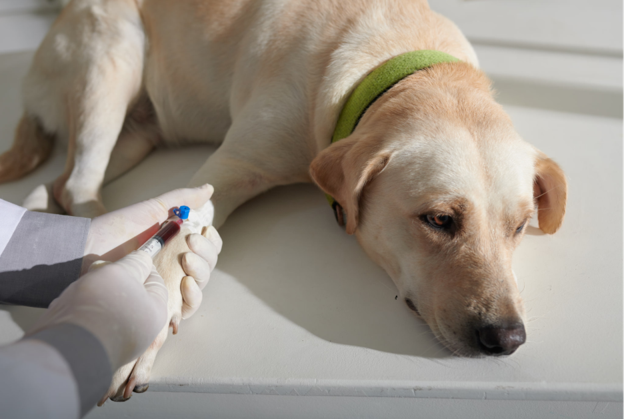
Early recognition of immune-mediated hemolytic anemia (IMHA) can significantly influence clinical decision-making and patient management. Although IMHA is a common autoimmune disorder in dogs, its clinical presentation is often nonspecific, making diagnosis challenging during the initial examination. Patients may present with signs attributable to anemia, hemolysis, or systemic inflammation, and the severity of clinical signs often depends on the rate at which anemia develops1. A thorough physical examination supported by hematological and biochemical evaluation remains essential for identifying affected dogs and determining the extent of disease involvement.
Recognizing the Clinical Presentation
The history and clinical examination provide valuable clues when IMHA is suspected. Dogs commonly present with signs related to reduced oxygen-carrying capacity and the body's compensatory response to anemia. In cases where anemia develops gradually, clinical abnormalities may remain subtle until the disease becomes severe1.
Common clinical findings include:
- Anorexia
- Lethargy
- Pale mucous membranes
- Jaundice (icterus)
- Hemoglobinuria resulting in port wine-colored urine
- Bilirubinuria
- Tachypnea
- Tachycardia
- Splenomegaly and hepatomegaly2
While splenic and hepatic macrophages are the principal sites of erythrocyte destruction, enlargement of these organs is not consistently observed in every patient1.
Jaundice is another important clinical finding. Severe anemia may impair hepatic function, reducing bilirubin clearance while increasing bilirubin production due to accelerated erythrocyte destruction. Icterus is usually first evident on the mucous membranes before becoming apparent on the skin as bilirubin concentrations continue to rise1.
Approximately 50% to 70% of dogs with IMHA may also have concurrent thrombocytopenia. When immune-mediated thrombocytopenia accompanies IMHA, the condition is referred to as Evans syndrome. These patients may exhibit bleeding manifestations such as epistaxis, petechiae, ecchymoses, and melena due to reduced circulating platelet numbers3.
Hematological Findings That Support Clinical Suspicion
Complete blood count (CBC) remains one of the most informative laboratory investigations in dogs with suspected IMHA. Findings typically reflect an acute hemolytic crisis together with evidence of regenerative anemia and tissue hypoxia.
A CBC commonly reveals:
- Normocytic, normochromic regenerative anemia
- Hematocrit ≤30%
- Polychromasia
- Anisocytosis
- Nucleated red blood cells1
Spherocytosis
Spherocytes are among the most characteristic hematological findings in canine IMHA. Partial removal of antibody-coated erythrocyte membranes by splenic or hepatic macrophages transforms normal biconcave erythrocytes into smaller, spherical cells lacking central pallor. Their presence is highly suggestive of extravascular hemolysis and has been identified in 89% to 95% of affected dogs1.
Reticulocytosis
Reticulocytosis reflects an active regenerative response to hemolysis. Increased circulating reticulocytes, together with nucleated RBCs, anisocytosis, and polychromasia, indicate that the bone marrow is responding appropriately to anemia1.
Leukocytosis
Many dogs develop an inflammatory leukogram characterized by neutrophilia with a left shift. In severe inflammatory states, a leukoerythroblastic pattern with numerous nucleated erythrocytes may also be observed. Marked leukocytosis may indicate significant tissue injury secondary to severe anemic hypoxia1.
Serum Biochemistry and Urinalysis
Biochemical abnormalities frequently accompany hematological changes and help assess the systemic impact of IMHA.
Common findings include:
- Increased serum bilirubin
- Elevated lactate dehydrogenase
- Increased blood urea nitrogen
- Increased creatinine
- Elevated ALT and AST
- Decreased haptoglobin concentration1
Hyperbilirubinemia results from increased bilirubin production following erythrocyte destruction, together with reduced hepatic clearance during severe anemia. In cases of intravascular hemolysis, circulating free hemoglobin binds to haptoglobin, resulting in reduced serum haptoglobin concentrations1.
Urinalysis further supports clinical assessment. Bilirubinuria is commonly associated with extravascular hemolysis, whereas hemoglobinuria is more suggestive of intravascular erythrocyte destruction1.
Bone Marrow Response1
Most dogs with IMHA demonstrate an active regenerative erythroid response. However, some patients may initially lack reticulocytosis because sufficient time has not elapsed for marrow regeneration to become evident. In prolonged non-regenerative cases, immune-mediated destruction of erythroid precursor cells may impair erythropoiesis despite persistent anemia.
Practical Clinical Insights
- Evaluate every anemic dog systematically for evidence of both hemolysis and regeneration rather than relying on a single laboratory finding.
- Pale mucous membranes, jaundice, hemoglobinuria, and lethargy together should heighten suspicion for IMHA and prompt further investigation.
- Careful examination of the peripheral blood smear for spherocytes remains an important component of routine hematological assessment.
- CBC, serum biochemistry, and urinalysis should be interpreted collectively, as each provides complementary information regarding disease severity and organ involvement.
- Concurrent thrombocytopenia should prompt consideration of Evans syndrome, particularly in dogs presenting with spontaneous bleeding.
Conclusion
Canine IMHA often presents with a combination of nonspecific clinical signs and characteristic hematobiochemical abnormalities. Careful assessment of clinical presentation, supported by CBC, blood smear evaluation, serum biochemistry, urinalysis, and bone marrow findings where appropriate, enables veterinarians to recognize affected patients promptly and determine the extent of systemic involvement. A structured interpretation of these findings forms the foundation for subsequent diagnostic confirmation and therapeutic planning.
References
- Karthik I, Dubey A, Swamy M, Verma Y, Jatav M, Khare A, Rakesh S. Immune mediated hemolytic anemia in dogs–an overview. Indian J Agric Allied Sci. 2022;8(4) https://www.researchgate.net/publication/377525778
- Manev, I. andMarincheva, V. (2018). Canine immune-mediated hemolytic anemia - brief review. Tradition and Modernity in Veterinary Medicine, 3(4): 59-64. https://www.researchgate.net/profile/Iliyan-Manev/publication/323959897
- Archer T, Mackin A. Diagnosis of immune-mediated hemolytic anemia. Today’s Veterinary Practice. 2013 Sep 11;7(8):32-6. https://todaysveterinarypractice.com/wp-content/uploads/sites/4/2016/09/T1307F04.pdf
Related Contents

Article
The Elephant is Walking Differently: Where Should the Investigation Begin?
When an elephant begins walking differently, the first challenge is not necessarily to identify the...
Article
When an Elephant’s Blood Report Says “Abnormal”: How Should It Be Interpreted?
A laboratory report can flag a value as abnormal even when an elephant appears clinically healthy. C...
.jpg)
Upcoming Event
World Animal Day
World Animal Day highlights the importance of protecting animal health and welfare through responsib...
Video
Rabies: Epitome of the One Health Paradigm
Rabies exemplifies the One Health approach, linking animal health, human health, veterinary services...
Video
One Health Webinar on Important Updates on Rabies for Veterinary Practitioners and Students
Rabies remains a major zoonotic disease requiring vigilance across veterinary practice and public he...
.jpg)
Video
Transjugular Removal of Canine Heartworms: A Clinical and Surgical Overview
Transjugular removal of canine heartworms is a specialized intervention used in selected dogs with s...

Article
Feline Pregnancy Endocrinology: Hormonal Changes and Placental Function
Pregnancy in cats involves a distinctive endocrine pattern in which ovarian and placental hormones w...

Article
Hormonal Control of Pregnancy Termination in Cats
Pregnancy maintenance in cats depends strongly on coordinated hormonal support, particularly progest...