Article
Chronic Diarrhoea in Adult Horses: Building a Practical Diagnostic Roadmap
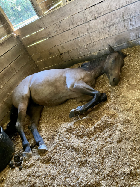
Chronic diarrhoea is a common yet challenging presentation in equine practice. Clinical signs are often non-specific, and definitive diagnosis may require a combination of laboratory testing, imaging, microbiological investigations, and histopathology1. A systematic diagnostic approach can help clinicians identify underlying causes and guide appropriate treatment strategies.
History Taking: The First Diagnostic Tool
A detailed and chronological history is critical when evaluating horses with chronic diarrhoea. Information regarding onset, duration, faecal consistency, seasonality, dietary changes, travel history, environmental factors, previous illnesses, and response to treatment can provide important diagnostic clues1.
Age may help narrow differential diagnoses. Larval cyathostominosis, one of the most important causes of chronic diarrhoea in horses, is more commonly reported in younger animals. Lawson et al. (2023) found that 92% of horses diagnosed with larval cyathostominosis presented with diarrhoea, with a median age of 2 years2.
Clinical signs frequently include weight loss, inappetence, and lethargy. Although less common, recurrent colic has been reported in approximately 19.6% of affected horses1.
Physical Examination: Looking Beyond Faecal Consistency
Interestingly, horses with chronic diarrhoea may have normal vital parameters despite longstanding diseases. Fever, when present, is more commonly associated with inflammatory disorders but may also occur as a paraneoplastic syndrome1.
Common examination findings include:
- Poor body condition and muscle wastage
- Ventral, mandibular, or limb oedema
- Increased gastrointestinal sounds
- Intestinal wall thickening or masses detected during rectal examination
Dermatological abnormalities should raise suspicion for multisystemic eosinophilic epitheliotropic disease (MEED). Approximately 77% of reported MEED cases present with dermatitis characterised by alopecia, hyperkeratosis, exudation, and lichenification affecting the face, limbs, and ventrum1.
Clinical Pathology: Hypoalbuminaemia as a Key Indicator
A complete blood count and serum biochemistry profile should be considered essential in every case.
The most consistent laboratory abnormality in horses with chronic diarrhoea is hypoproteinaemia, particularly hypoalbuminaemia, reflecting protein loss through the gastrointestinal tract1.
Additional findings may include1,3:
- Anaemia due to malabsorption, blood loss, or chronic inflammation
- Neutrophilia or neutropenia in granulomatous enteritis (GE)
- Eosinophilia in selected eosinophilic gastrointestinal disorders
- Electrolyte abnormalities such as hyponatraemia, hypokalaemia, hypochloraemia, and hypocalcaemia in severe cases
Hypoalbuminaemia has been reported in 75% of horses with lymphocytic-plasmacytic enteritis and is a consistent finding in horses with cyathostominosis2.
Elevated liver enzymes may also provide diagnostic clues. Increased gamma-glutamyl transferase (GGT) concentrations were observed in 73% of horses with MEED but not in horses with granulomatous enteritis, suggesting a potential differentiating marker1.
Faecal Diagnostics: More Than Just Egg Counts
Comprehensive faecal examination remains a cornerstone of investigation.
Recommended tests include:
- Faecal flotation
- Wet smear examination for larvae
- Sand sedimentation testing
- Bacterial culture
- Molecular diagnostic panels
Larval cyathostominosis remains one of the most common causes of chronic diarrhoea in adult horses. However, clinicians should recognise that faecal egg counts do not correlate well with parasite burden, and negative results do not exclude disease1. Faecal PCR testing is particularly useful when Lawsonia intracellularis infection is suspected in younger horses. Combining PCR with serology improves diagnostic sensitivity.
Sand enteropathy should also be considered, particularly in horses grazing sandy paddocks. Faecal sedimentation testing has demonstrated good diagnostic utility, with a reported sensitivity of 83% when compared with radiography4.
Ultrasonography: A High-Yield Imaging Modality
Transabdominal ultrasonography is one of the most valuable diagnostic tools available for investigating chronic diarrhoea.
Potential findings include:
- Thickened intestinal walls
- Altered intestinal motility
- Right dorsal colon abnormalities
- Sand accumulation
- Abdominal masses or abscesses
Diffuse intestinal wall thickening is commonly reported in infiltrative bowel diseases and intestinal lymphoma. Ultrasonographic thickening of the right dorsal colon has been documented in 77% of horses with right dorsal colitis1.
Marked caecal and colonic wall thickening may also support a diagnosis of larval cyathostominosis.
Biopsy and Functional Testing
Rectal mucosal biopsy remains a useful and relatively accessible diagnostic procedure, with reported diagnostic accuracy ranging from 50% to 82%5.
When malabsorption is suspected, oral glucose absorption tests (OGAT) and d-xylose absorption tests can help evaluate small intestinal absorptive function. Abnormal absorption test results were reported in 75% of horses diagnosed with lymphocytic-plasmacytic enteritis1.
For selected cases, particularly when infiltrative bowel disease or neoplasia is strongly suspected, full-thickness intestinal biopsies obtained via laparotomy or laparoscopy may provide the highest diagnostic yield6.
Clinical Take-Home Points
- Chronic diarrhoea requires a structured and systematic diagnostic approach.
- Young horses with diarrhoea and hypoalbuminaemia should always prompt consideration of larval cyathostominosis.
- Hypoalbuminaemia remains one of the most consistent laboratory findings across many chronic enteropathies.
- Ultrasonography is invaluable for identifying infiltrative intestinal disease, right dorsal colitis, and sand accumulation.
- Faecal egg counts alone are insufficient to exclude cyathostomin disease.
- Rectal biopsy, absorption testing, and full-thickness intestinal biopsy can significantly improve diagnostic accuracy in complex cases.
Early recognition and a logical diagnostic pathway improve the likelihood of identifying treatable causes of chronic diarrhoea and optimising long-term outcomes for equine patients.
References
- Sjolin E, Lack A, Arroyo LG. Diagnostic approach to chronic diarrhoea in adult horses. Equine Veterinary Education. 2025 Jun;37(6):328-36. https://beva.onlinelibrary.wiley.com/doi/pdf/10.1111/eve.14062
- Lawson AL, Malalana F, Mair TS. Larval cyathostominosis: Clinicopathological data and treatment outcomes of 38 hospitalised horses (2009–2020). Equine veterinary education. 2023 Aug;35(8):424-35. https://doi.org/10.1111/eve.13782
- Villagrán CC, Vogt D, Gupta A, Fernández EA. Inflammatory bowel disease characterized by multisystemic eosinophilic epitheliotropic disease (MEED) in a horse in Saskatchewan, Canada. The Canadian Veterinary Journal. 2021 Nov;62(11):1190. https://pmc.ncbi.nlm.nih.gov/articles/PMC8543654/
- Hukkinen V. The diagnostic accuracy of the plastic glove test for diagnosis of sand enteropathy in the horse. Licenciate thesis, University of Helsinki, Faculty of Veterinary Medicine: Department of Equine and Small Animal Medicine. 2015 Apr. https://helda.helsinki.fi/server/api/core/bitstreams/0a21fc25-3501-4aa4-9182-8b5470bac105/content
- Boshuizen B, Ploeg M, Dewulf J, Klooster S, Bruijn MD, Picavet MT, Palmers K, Plancke L, Cock HD, Theelen M, Delesalle C. Inflammatory bowel disease (IBD) in horses: a retrospective study exploring the value of different diagnostic approaches. BMC veterinary research. 2018 Jan 19;14(1):21. https://link.springer.com/article/10.1186/s12917-018-1343-1
- Hostetter JM, Uzal FA. Gastrointestinal biopsy in the horse: overview of collection, interpretation, and applications. Journal of Veterinary Diagnostic Investigation. 2022 May;34(3):376-88. https://doi.org/10.1177/10406387221085584
Related Contents

Upcoming Event
Homeopathy in Pet Animal Practice
Homeopathy continues to be used by some veterinarians and pet owners as a complementary approach in...

Upcoming Event
Advanced Veterinary Transfusion Medicine
Transfusion medicine has become an essential component of modern veterinary critical care and intern...

Upcoming Event
Effect of Heat Stress on Bovine Reproduction
Heat stress is a major challenge in cattle production systems, particularly in regions with high tem...

Upcoming Event
Lumpy Skin Disease: From Signs to Field level control
Lumpy Skin Disease (LSD) has emerged as a significant transboundary viral disease affecting cattle,...
.jpg)
Upcoming Event
Hemogram with Special Reference to IMHA
Anaemia is a common clinical finding in canine and feline practice and may result from blood loss, h...

Upcoming Event
One Health in Action to Combat Zoonotic Diseases
Zoonotic diseases continue to pose significant challenges to global health, animal health, and envir...

Article
PRP, IRAP or Stem Cells? Choosing the Right Biologic for Equine Osteoarthritis
Biologics are everywhere—but which one to choose? Regenerative...

Article
Beyond Wear and Tear: Understanding How Osteoarthritis Develops in Performance Horses
For equine athletes, peak performance and joint health exist in a delicate balance. Whether it is a...