Article
Metabolomics in Equine Osteoarthritis: A New Frontier for Early Diagnosis
Osteoarthritis (OA) remains one of the leading causes of lameness and reduced athletic performance in sport horses. Despite advances in imaging and clinical evaluation, early diagnosis continues to be challenging, often delaying intervention until irreversible joint damage has occurred1.
Why Early Detection Matters
OA is a complex disease involving cartilage degradation, synovial inflammation, subchondral bone remodeling, and progressive loss of joint function. Inflammatory mediators released within the joint—including cytokines, prostaglandins, and nitric oxide—create a vicious cycle that accelerates tissue damage2,3.
Current treatment strategies focus largely on symptom management through anti-inflammatory medications and intra-articular therapies, rather than disease modification1. This has intensified the search for biomarkers capable of identifying OA before significant structural changes become evident.
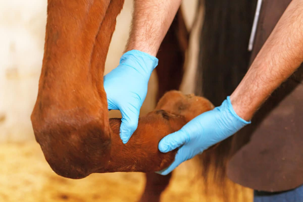
Synovial Fluid: A Window into Joint Health
Because synovial fluid (SF) is in direct contact with articular tissues, it provides valuable insights into ongoing pathological processes within the joint. Traditional assessments such as total protein concentration and white blood cell counts are useful but often lack sensitivity for early OA detection3.
Recent advances in metabolomics—the study of small molecules produced during cellular metabolism—have opened new possibilities for equine OA diagnostics4.
What Metabolomics Reveals
Using advanced analytical techniques such as proton nuclear magnetic resonance (^1H-NMR) spectroscopy, researchers have identified distinct metabolic signatures associated with OA5,6.
Several metabolites were found to be significantly altered in OA-affected joints:
- Tryptophan concentrations decrease during OA and may reflect inflammatory changes within the synovium1,7.
- Phenylalanine and tyrosine, important amino acids involved in cellular regulation, are reduced in diseased joints5.
- Glycine, a critical component of collagen synthesis, is significantly lower, potentially reflecting ongoing cartilage degeneration1.
- Glutamine levels decrease, suggesting increased utilization during inflammatory and tissue repair processes8,9.
- Arginine concentrations decline, supporting evidence that altered nitric oxide metabolism and oxidative stress contribute to OA development10,11.
- Methionine, known for its antioxidant properties, is also reduced, possibly due to increased consumption during chronic inflammation12.
Interestingly, 1,3-dihydroxyacetone was found at higher concentrations in OA joints. Researchers speculate that this may represent an adaptive response by chondrocytes attempting to preserve cartilage integrity and glycosaminoglycan content [41–43].
What This Means for Practising Veterinarians
Although metabolomic testing is not yet part of routine equine practice, the findings are highly promising. Identifying metabolic changes before radiographic lesions become apparent could enable:
- Earlier OA diagnosis
- Improved monitoring of disease progression
- Better assessment of treatment response
- More targeted management strategies for athletic horses
Looking Ahead
Metabolomics is transforming our understanding of equine joint disease. As research continues, synovial fluid biomarkers may become valuable tools alongside imaging and clinical examination, helping veterinarians detect OA sooner and intervene before performance-limiting damage occurs.
Key Clinical Takeaway
Metabolomic profiling of synovial fluid has identified several biomarkers associated with equine OA. While not yet a routine diagnostic tool, this technology has significant potential to improve early detection and long-term management of joint disease in sport horses.
References
- Laus F, Gialletti R, Bazzano M, Laghi L, Dini F, Marchegiani A. Synovial fluid metabolome can differentiate between healthy joints and joints affected by osteoarthritis in horses. Metabolites. 2023 Aug 4;13(8):913. https://doi.org/10.3390/metabo13080913
- Menarim BC, Gillis KH, Oliver A, Ngo Y, Werre SR, Barrett SH, Rodgerson DH, Dahlgren LA. Macrophage activation in the synovium of healthy and osteoarthritic equine joints. Frontiers in Veterinary Science. 2020 Nov 26;7:568756. https://www.frontiersin.org/journals/veterinary-science/articles/10.3389/fvets.2020.568756/pdf
- Ribitsch I, Oreff GL, Jenner F. Regenerative medicine for equine musculoskeletal diseases. Animals. 2021 Jan 19;11(1):234. https://doi.org/10.3390/ani11010234
- Bujak R, Struck-Lewicka W, Markuszewski MJ, Kaliszan R. Metabolomics for laboratory diagnostics. Journal of pharmaceutical and biomedical analysis. 2015 Sep 10;113:108-20. https://www.academia.edu/download/39943849/Metabolomics_for_laboratory_diagnostics20151112-11019-l4rqui.pdf
- Anderson JR, Phelan MM, Clegg PD, Peffers MJ, Rubio-Martinez LM. Synovial fluid metabolites differentiate between septic and nonseptic joint pathologies. Journal of proteome research. 2018 Jul 3;17(8):2735-43. https://pubs.acs.org/doi/pdf/10.1021/acs.jproteome.8b00190
- Graham RJ, Anderson JR, Phelan MM, Cillan‐Garcia E, Bladon BM, Taylor SE. Metabolomic analysis of synovial fluid from Thoroughbred racehorses diagnosed with palmar osteochondral disease using magnetic resonance imaging. Equine veterinary journal. 2020 May;52(3):384-90. https://livrepository.liverpool.ac.uk/3066643/1/Graham%20et%20al.%2C%202019%20-%20Accepted%20Article.pdf
- Nowicka-Stążka P, Langner E, Turski W, Rzeski W, Parada-Turska J. Quinaldic acid in synovial fluid of patients with rheumatoid arthritis and osteoarthritis and its effect on synoviocytes in vitro. Pharmacological Reports. 2018 Mar;70(2):277-83. https://www.researchgate.net/profile/Wojciech-Rzeski/publication/320199542
- Kim H. Glutamine as an immunonutrient. Yonsei medical journal. 2011 Oct 20;52(6):892. https://synapse.koreamed.org/pdf/10.3349/ymj.2011.52.6.892
- Takahashi S, Saegusa J, Sendo S, Okano T, Akashi K, Irino Y, Morinobu A. Glutaminase 1 plays a key role in the cell growth of fibroblast-like synoviocytes in rheumatoid arthritis. Arthritis research & therapy. 2017 Apr 11;19(1):76. https://link.springer.com/content/pdf/10.1186/s13075-017-1283-3.pdf
- McNeal CJ, Meininger CJ, Reddy D, Wilborn CD, Wu G. Safety and effectiveness of arginine in adults. The Journal of nutrition. 2016 Dec 1;146(12):2587S-93S. https://academic.oup.com/jn/article-pdf/146/12/2587S/30020600/jn234740.pdf
- Carlson AK, Rawle RA, Wallace CW, Adams E, Greenwood MC, Bothner B, June RK. Global metabolomic profiling of human synovial fluid for rheumatoid arthritis biomarkers. Clin Exp Rheumatol. 2019 May 1;37(3):393-9. https://www.clinexprheumatol.org/article.asp?a=12929
- Aledo JC. Methionine in proteins: The Cinderella of the proteinogenic amino acids. Protein Science. 2019 Oct;28(10):1785-96. https://pmc.ncbi.nlm.nih.gov/articles/PMC6739822/pdf/PRO-28-1785.pdf
Related Contents

Upcoming Event
Homeopathy in Pet Animal Practice
Homeopathy continues to be used by some veterinarians and pet owners as a complementary approach in...

Upcoming Event
Advanced Veterinary Transfusion Medicine
Transfusion medicine has become an essential component of modern veterinary critical care and intern...

Upcoming Event
Effect of Heat Stress on Bovine Reproduction
Heat stress is a major challenge in cattle production systems, particularly in regions with high tem...

Upcoming Event
Lumpy Skin Disease: From Signs to Field level control
Lumpy Skin Disease (LSD) has emerged as a significant transboundary viral disease affecting cattle,...
.jpg)
Upcoming Event
Hemogram with Special Reference to IMHA
Anaemia is a common clinical finding in canine and feline practice and may result from blood loss, h...

Upcoming Event
One Health in Action to Combat Zoonotic Diseases
Zoonotic diseases continue to pose significant challenges to global health, animal health, and envir...

Article
PRP, IRAP or Stem Cells? Choosing the Right Biologic for Equine Osteoarthritis
Biologics are everywhere—but which one to choose? Regenerative...

Article
Beyond Wear and Tear: Understanding How Osteoarthritis Develops in Performance Horses
For equine athletes, peak performance and joint health exist in a delicate balance. Whether it is a...