Article
Ear Cytology Made Practical: What Every Veterinarian Should Look for Before Starting Treatment
Empirical treatment for canine otitis without performing ear cytology is still common in clinical practice.
Ear cytology is inexpensive, quick, and arguably the single most valuable diagnostic test for otitis externa. It not only identifies the microorganisms present but also guides treatment selection, monitors therapeutic response, and supports responsible antimicrobial use1,2,3,4.
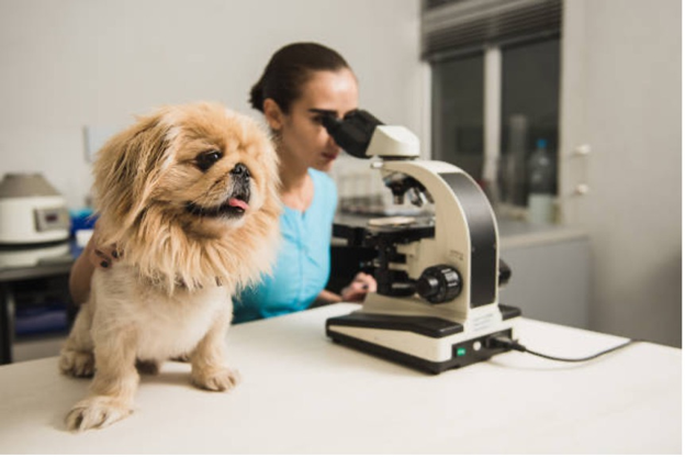
Before reaching for an ear cleaner or topical medication, spend a few minutes looking down the microscope—it can completely change your treatment plan.
Why Every Otitis Consultation Should Include Cytology
Clinical signs such as head shaking, pruritus, erythema or ear discharge tell you that inflammation is present—but they do not reveal its cause.
Ear cytology helps answer several important clinical questions:
- Is the infection bacterial, fungal or mixed?
- Are cocci or rods predominating?
- Is Malassezia contributing to the inflammation?
- Is there evidence of marked inflammation?
- Is the patient responding to treatment at follow-up?
Without cytology, treatment becomes empirical rather than evidence-based1.
Collect the Sample Correctly
A good cytology begins with a good sample.
For most cases:
- Sample the horizontal ear canal, where microorganisms are most representative.
- Use a sterile cotton swab or cytology loop.
- Roll—not smear—the material gently onto a clean microscope slide.
- Allow the slide to air dry before staining.
- Diff-Quik remains the most commonly used stain in clinical practice.
Whenever possible, collect the sample before ear cleaning or administering medication, as both can reduce the diagnostic yield.
What Should You Look For?
Rather than trying to identify every organism, focus on the findings that influence clinical decisions.
1. Bacteria
The first question is whether bacteria are present.
Then determine whether they are predominantly:
Cocci
These are commonly associated with Staphylococcus pseudintermedius and other Gram-positive organisms. Many uncomplicated cases respond well to appropriate topical therapy (1).
Rods (Bacilli)
Rod-shaped bacteria should immediately raise concern for more complicated disease.
Persistent rod infections may indicate organisms such as Pseudomonas aeruginosa or Proteus species, particularly in chronic or previously treated ears. These patients often require bacterial culture and susceptibility testing before treatment is adjusted2,3.
2. Malassezia
Malassezia pachydermatis appears as characteristic peanut- or footprint-shaped budding yeast.
Small numbers may occasionally be present in healthy ears.
However, large numbers together with pruritus, erythema and ceruminous discharge strongly suggest clinically significant yeast overgrowth1.
Remember that Malassezia is frequently secondary to underlying allergic skin disease rather than being the primary problem.
3. Inflammatory Cells
Neutrophils provide useful information about disease activity.
Large numbers of degenerate neutrophils suggest active bacterial infection.
Conversely, abundant microorganisms with minimal inflammatory cells may indicate colonisation rather than severe infection, emphasising the need to interpret cytology alongside the patient's clinical signs.
4. Mixed Populations
Finding both bacteria and yeast on the same slide is not uncommon.
Mixed infections are frequently encountered in chronic otitis and generally require combination topical therapy together with investigation of the underlying primary disease.
Simply targeting one organism may result in incomplete resolution.
Cytology Should Guide Treatment Decisions
The cytology findings should directly influence the next clinical step.
|
Cytology finding |
Clinical implication |
|
Predominantly cocci |
Appropriate topical antibacterial therapy may be sufficient in uncomplicated cases. |
|
Predominantly rods |
Consider chronic infection, obtain bacterial culture if indicated, and reassess treatment strategy. |
|
Predominantly Malassezia |
Select an appropriate antifungal-containing topical product and investigate underlying allergy. |
|
Mixed bacteria and yeast |
Combination topical therapy and investigation for primary disease are often required. |
|
Minimal organisms despite severe inflammation |
Reconsider differential diagnoses, including allergic or inflammatory disease. |
Cytology Doesn't Replace Culture
One common misconception is that cytology eliminates the need for bacterial culture.
In reality, the two tests answer different questions.
Cytology tells you:
- What organisms are present today.
- Whether treatment appears to be working.
Culture tells you:
- Which bacteria are growing.
- Their antimicrobial susceptibility.
- Which antibiotic is most appropriate for complicated infections2.
Culture is particularly valuable for:
- Chronic otitis
- Recurrent infections
- Rod-shaped bacterial infections
- Poor response to empirical therapy
- Suspected multidrug-resistant organisms
Repeat Cytology at Follow-Up
One of the most overlooked steps in otitis management is repeat cytology.
Clinical improvement does not necessarily indicate microbiological resolution.
A dog may stop scratching while significant bacterial or yeast populations remain within the ear canal.
Repeat cytology helps determine whether:
- Infection has resolved.
- Treatment should continue.
- Medication can safely be discontinued.
- The apparent "recurrence" is actually an incomplete resolution of the original episode.
Common Cytology Mistakes
Avoid these common pitfalls:
- Collecting samples after ear cleaning.
- Sampling only superficial debris.
- Treating without performing cytology.
- Interpreting microorganisms without considering clinical signs.
- Failing to repeat cytology before stopping treatment.
- Ignoring rod-shaped bacteria in recurrent cases.
Clinical Pearl
Ear cytology should not be viewed as an optional diagnostic test—it should become a routine part of every otitis consultation.
A five-minute examination under the microscope often provides more clinically useful information than changing medications based on appearance alone.
Key Takeaways
- Ear cytology is the cornerstone of diagnosing and monitoring canine otitis externa.
- Always collect samples before cleaning or medicating the ear.
- Differentiate cocci, rods, yeast and inflammatory cells, as each influences treatment decisions.
- Rod-shaped bacteria and recurrent infections should prompt consideration of bacterial culture and susceptibility testing.
- Repeat cytology at follow-up confirms microbiological resolution and helps reduce recurrence.
References
- Bajwa J. Canine otitis externa—Treatment and complications. Canadian Veterinary Journal. 2019;60(1):98–101. PMCID: PMC6294027. Available from: https://pmc.ncbi.nlm.nih.gov/articles/PMC6294027/
- Leonard, C.; Thiry, D.; Taminiau, B.; Daube, G.; Fontaine, J. External Ear Canal Evaluation in Dogs with Chronic Suppurative Otitis Externa: Comparison of Direct Cytology, Bacterial Culture and 16S Amplicon Profiling. Vet. Sci. 2022, 9, 366. https://doi.org/10.3390/vetsci9070366
- Secker B, Shaw S, Atterbury RJ. Pseudomonas spp. in Canine Otitis Externa. Microorganisms. 2023;11(11):2650. DOI: 10.3390/microorganisms11112650. PMCID: PMC10671989. https://www.mdpi.com/2076-2607/11/11/2650
- Ponn PC, Tipold A, Volk AV, et al. Can We Minimize the Risk of Dogs Developing Canine Otitis Externa? Animals. 2024;14(17):2537. DOI: 10.3390/ani14172537. PMCID: PMC11394639. Available from: https://pmc.ncbi.nlm.nih.gov/articles/PMC11394639/
Related Contents

Upcoming Event
African Swine Fever: Diagnosis and Management
African swine fever can cause severe disease and significant losses in affected pig populations.&nbs...
.jpg)
Video
Transjugular Removal of Canine Heartworms: A Clinical and Surgical Overview
Transjugular removal of canine heartworms is a specialized intervention used in selected dogs with s...

Article
Feline Pregnancy Endocrinology: Hormonal Changes and Placental Function
Pregnancy in cats involves a distinctive endocrine pattern in which ovarian and placental hormones w...

Article
Hormonal Control of Pregnancy Termination in Cats
Pregnancy maintenance in cats depends strongly on coordinated hormonal support, particularly progest...

Article
Feline Embryo Implantation and Early Placental Development
Pregnancy establishment in cats involves a sequence of precisely timed events between fertilization,...

Article
Ultrasonographic Monitoring of Pregnancy in Cats: A Gestational Timeline
Ultrasonography provides a practical way to follow the progression of feline pregnancy by identifyin...

Article
Feline Mismating: Pregnancy Diagnosis and Clinical Management
Mismating or unplanned mating is a common clinical problem in sexually mature queen cats because of...

Article
Medical Termination of Pregnancy in Queens: Drugs, Timing and Treatment Protocols
Medical termination of pregnancy (MTP) in queen cats should be approached according to the timing of...