Article
Diagnosing Glässer's Disease in Swine: A Practical Guide for Field Veterinarians
Glässer's disease remains an important cause of morbidity and mortality in nursery and early growing pigs, with significant consequences for productivity and herd profitability. Because the disease shares clinical and pathological features with several other bacterial infections, an accurate diagnosis requires more than recognizing compatible clinical signs. A systematic approach that combines clinical evaluation, postmortem examination, appropriate sample collection, and laboratory confirmation allows veterinarians to identify pathogenic Glaesserella (Haemophilus) parasuis and make informed treatment and herd management decisions.
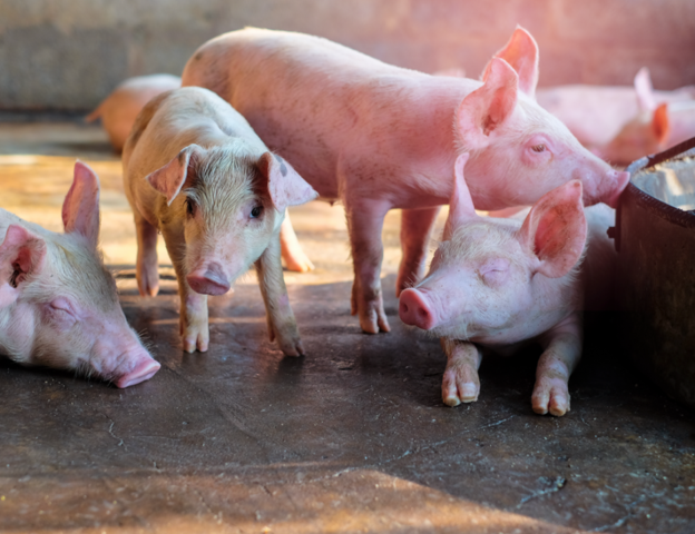
Recognizing Clinical Signs in the Field
The diagnostic process begins with careful observation of affected pigs. Clinical signs are not specific and should always be interpreted alongside the herd history and disease pattern.
Common clinical findings include:
- Fever
- Cough
- Abdominal breathing
- Swollen joints
- Lameness
- Central nervous system signs, including lateral decubitus, pedaling movements and tremors1.
Acute cases typically occur after weaning, when stress associated with management changes coincides with declining maternal immunity, creating favourable conditions for disease development1.
Postmortem Findings Support Clinical Suspicion
Necropsy provides valuable diagnostic information, particularly in acute outbreaks. Piglets commonly present with fibrinous polyserositis, while purulent catarrhal pneumonia may also be observed in some animals1. Chronically affected pigs may exhibit reduced growth together with persistent fibrinous polyserositis lesions at necropsy1.
Although these findings strongly support a diagnosis of Glässer's disease, similar lesions can also occur with infections caused by Streptococcus suis and Mycoplasma hyorhinis. Consequently, clinical and pathological observations should always be confirmed by laboratory testing before establishing the diagnosis1.
Laboratory Confirmation
Laboratory diagnosis is commonly based on bacterial isolation and PCR1. PCR offers a rapid method for detecting G. parasuis, whereas bacterial isolation remains essential whenever additional characterization of the isolate is required, particularly antimicrobial susceptibility testing.
An important consideration during interpretation is that G. parasuis is a heterogeneous species comprising both virulent and non-virulent strains. Therefore, identifying the bacterium alone does not necessarily confirm that it is responsible for clinical disease. Molecular diagnostic methods capable of determining serovar and predicting pathogenic potential have become valuable additions to routine diagnostics2,3,4.
Collecting the Right Samples
Appropriate sample collection greatly improves diagnostic success. Whenever possible, samples should be collected from several untreated pigs showing clear clinical signs. If systemic disease is suspected, specimens should include affected serosal surfaces, joints and the brain.
Respiratory samples require careful interpretation because colonizing strains may be aspirated into the lungs during euthanasia. For this reason, lung samples should only be collected when pneumonia is present and should be obtained from distal lung tissue5.
Strict aseptic sampling procedures are essential to minimise contamination. Samples should be transported rapidly under refrigeration (4–8 °C), and swabs should be placed in Amies transport medium to maintain bacterial viability5.
Understanding Molecular Diagnosis
Isolation of G. parasuis from systemic lesions is considered evidence of virulence because systemic dissemination is a recognised virulence characteristic. In contrast, recovery of the organism from the respiratory tract alone should be interpreted cautiously, since both virulent and non-virulent strains may colonize healthy pigs5.
Molecular serotyping has become an important tool for determining circulating serovars and may assist vaccine selection3. In addition, PCR assays targeting virulence-associated genes, including leader sequences of the vtaA genes, improve the ability to differentiate strains with higher pathogenic potential4. Multiplex PCR assays designed to detect sets of virulence-associated genes may also help identify pigs carrying virulent strains and assess the risk of disease development within a herd5.
Practical Clinical Insights
Successful diagnosis of Glässer's disease depends on integrating field observations with laboratory evidence. Examine clinically affected pigs early in the outbreak, collect samples before antimicrobial treatment whenever possible, and submit multiple tissues from untreated animals. Combine bacterial isolation with PCR to improve diagnostic confidence, and request antimicrobial susceptibility testing when treatment decisions are required. Interpreting laboratory results alongside lesion distribution, sampling site and the pathogenic potential of the detected strain enables more accurate diagnosis and supports more effective herd-level control strategies.
References
- Aragon V, Segalés J, Tucker AW. Glässer's disease. Diseases of swine. 2019 Jun 3:844-53. https://repositori.irta.cat/bitstream/handle/20.500.12327/3798/Aragon_Glasser_Anaporc_2024.pdf?sequence=1&isAllowed=y
- Howell KJ, Peters SE, Wang J, Hernandez-Garcia J, Weinert LA, Luan SL, Chaudhuri RR, Angen Ø, Aragon V, Williamson SM, Parkhill J. Development of a multiplex PCR assay for rapid molecular serotyping of Haemophilus parasuis. Journal of clinical microbiology. 2015 Dec;53(12):3812-21. https://journals.asm.org/doi/pdf/10.1128/jcm.01991-15
- Galofré-Milà N, Correa-Fiz F, Lacouture S, Gottschalk M, Strutzberg-Minder K, Bensaid A, Pina-Pedrero S, Aragon V. A robust PCR for the differentiation of potential virulent strains of Haemophilus parasuis. BMC veterinary research. 2017 May 8;13(1):124. https://link.springer.com/content/pdf/10.1186/s12917-017-1041-4.pdf
- Howell KJ, Weinert LA, Peters SE, Wang J, Hernandez-Garcia J, Chaudhuri RR, Luan SL, Angen Ø, Aragon V, Williamson SM, Langford PR. “Pathotyping” multiplex PCR assay for Haemophilus parasuis: a tool for prediction of virulence. Journal of Clinical Microbiology. 2017 Sep;55(9):2617-28. https://journals.asm.org/doi/pdf/10.1128/jcm.02464-16
- Costa-Hurtado M, Barba-Vidal E, Maldonado J, Aragon V. Update on Glässer’s disease: How to control the disease under restrictive use of antimicrobials. Veterinary microbiology. 2020 Mar 1;242:108595. https://www.sciencedirect.com/science/article/pii/S0378113519314713
Related Contents

Article
When an Elephant’s Blood Report Says “Abnormal”: How Should It Be Interpreted?
A laboratory report can flag a value as abnormal even when an elephant appears clinically healthy. C...
.jpg)
Upcoming Event
World Animal Day
World Animal Day highlights the importance of protecting animal health and welfare through responsib...
Video
Rabies: Epitome of the One Health Paradigm
Rabies exemplifies the One Health approach, linking animal health, human health, veterinary services...
Video
One Health Webinar on Important Updates on Rabies for Veterinary Practitioners and Students
Rabies remains a major zoonotic disease requiring vigilance across veterinary practice and public he...
.jpg)
Video
Transjugular Removal of Canine Heartworms: A Clinical and Surgical Overview
Transjugular removal of canine heartworms is a specialized intervention used in selected dogs with s...

Article
Feline Pregnancy Endocrinology: Hormonal Changes and Placental Function
Pregnancy in cats involves a distinctive endocrine pattern in which ovarian and placental hormones w...

Article
Hormonal Control of Pregnancy Termination in Cats
Pregnancy maintenance in cats depends strongly on coordinated hormonal support, particularly progest...

Article
Feline Embryo Implantation and Early Placental Development
Pregnancy establishment in cats involves a sequence of precisely timed events between fertilization,...