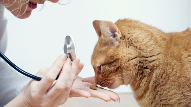
Article
Not Every Feline Urolith Needs Surgery: Choosing the Right Treatment Strategy
Managing feline urolithiasis extends beyond identifying urinary stones—it requires selecting the most appropriate treatment for the individual patient. While surgery is often considered the default option, not every urolith requires immediate surgical removal. Stone composition, location, size, degree of obstruction and the cat's overall clinical condition all influence treatment decisions. An evidence-based approach helps avoid unnecessary procedures while ensuring timely intervention when required¹˒².
Begin with Patient Stabilisation
Before deciding how to remove a stone, clinicians should determine whether the cat is stable. Cats presenting with urethral obstruction, severe dehydration, electrolyte abnormalities or post-renal azotemia require immediate stabilisation before definitive treatment is attempted².
Assessment should include:
- Hydration status
- Serum electrolytes
- Blood urea nitrogen (BUN) and creatinine
- Pain assessment
- Bladder size and integrity
Failure to correct metabolic abnormalities before intervention may increase anaesthetic and surgical risk².
Clinical Pearl: Treat the patient before treating the stone.
Identify the Stone Before Choosing the Treatment
Treatment should never be based solely on the presence of a urinary stone. Whenever possible, clinicians should predict stone composition using signalment, urinalysis, imaging characteristics and clinical history before selecting therapy¹.
This distinction is critical because:
- Struvite uroliths may respond to therapeutic dissolution diets under appropriate clinical conditions¹.
- Calcium oxalate uroliths do not dissolve medically and generally require physical removal¹.
Attempting dietary dissolution of calcium oxalate stones delays definitive treatment and may increase the risk of persistent clinical signs or obstruction.
When Is Medical Management Appropriate?
Medical dissolution is most successful when:
- Struvite urolithiasis is strongly suspected.
- There is no urethral obstruction.
- The patient is clinically stable.
- Owners are likely to comply with dietary recommendations and follow-up imaging¹.
During dissolution therapy, repeat radiographic evaluation is essential to confirm reduction in stone size and ensure treatment remains effective¹.
Clinical Pearl: Medical dissolution requires monitoring—not simply changing the diet.
When Surgery Is the Better Option
Surgical intervention remains the treatment of choice for:
- Calcium oxalate uroliths.
- Large bladder stones unlikely to dissolve.
- Persistent clinical signs despite medical management.
- Recurrent urinary obstruction.
- Cases where stone composition is uncertain and immediate removal is indicated¹˒².
Cystotomy continues to be the most commonly performed procedure for bladder urolith removal and provides the added advantage of recovering intact calculi for quantitative stone analysis².
Managing Urethral Uroliths
Urethral calculi require rapid intervention because complete obstruction can quickly become life-threatening. Initial management often involves careful catheterisation with retrograde urohydropropulsion, allowing stones to be flushed back into the bladder before cystotomy².
If urohydropropulsion is unsuccessful or catheterisation cannot be achieved, urethrotomy or referral for advanced intervention may be necessary².
Clinical Pearl: Whenever feasible, relocate urethral stones into the bladder rather than attempting direct urethral removal.
Stone Removal Is Only Half the Treatment
Successful surgery does not eliminate the risk of recurrence. Every retrieved urolith should be submitted for quantitative analysis because prevention strategies depend on knowing the stone's mineral composition¹.
Long-term management should include:
- Therapeutic nutrition tailored to stone type.
- Encouraging increased water intake.
- Periodic urinalysis.
- Follow-up imaging to detect early recurrence.
- Investigation of metabolic disorders such as hypercalcaemia in cats with calcium oxalate urolithiasis¹.
Failure to identify and address underlying risk factors increases the likelihood of recurrent disease.
Take-Home Message
There is no universal treatment for feline urolithiasis. The best outcomes are achieved when clinicians individualise therapy based on stone composition, anatomical location and the patient's clinical status. While medical dissolution is an excellent option for selected struvite stones, calcium oxalate calculi and obstructive uroliths generally require surgical intervention. Regardless of the treatment chosen, stone analysis and long-term preventive strategies remain essential to minimise recurrence and improve patient outcomes.
References
- Bartges JW. Feline calcium oxalate urolithiasis: risk factors and rational treatment approaches. Journal of Feline medicine and Surgery. 2016 Sep;18(9):712-22. https://journals.sagepub.com/doi/full/10.1177/1098612x16660442
- Yaygıngül R. Clinical, laboratory, radiography and ultrasonography findings and surgical treatment the lower urinary system urolithiasis in cats and dogs. Animal Health Production and Hygiene. 2024 Jun 6;13(1):23-30. https://dergipark.org.tr/en/download/article-file/3472658
Related Contents

Upcoming Event
ECG Interpretation Made Easy for Small Animal Practitioners
Electrocardiography (ECG) is an essential diagnostic tool in small animal practice, yet many clinici...

Upcoming Event
Positive Inotropes and Their Role in Cardiology
Positive inotropic agents are commonly used in cardiology to improve myocardial contractility and su...

Upcoming Event
Lesion-Based Diagnosis of Economically Important Poultry Diseases: A Visual Journey Through Gross Pathology
Accurate recognition of gross pathological lesions is essential for the diagnosis and control of pou...
.jpg)
Upcoming Event
Otitis in Cats and Dogs
Otitis is one of the most common ear disorders affecting the health and comfort of cats and dogs. Ga...
Upcoming Event
Postpartum Reproductive Disorders in Dairy Cattle
Postpartum reproductive disorders are a major cause of reduced fertility and economic losses in dair...
.jpg)
Upcoming Event
Bird Flu: A Bird's-Eye View
Avian influenza, commonly known as bird flu, remains one of the most significant infectious diseases...
.jpg)
Upcoming Event
Bovine Tuberculosis: Diagnostic Challenges and Pathological Features
Bovine tuberculosis is a chronic infectious disease that continues to impact cattle health, farm eco...

Upcoming Event
Post-Mortem Examination: Practical Tips for Field Veterinarians
Post-mortem examination is a valuable diagnostic tool that helps veterinarians determine the cause o...