Article
When Should You Suspect Urolithiasis in Cats? A Practical Diagnostic Approach for Veterinary Practice
Lower urinary tract signs are among the most frequent reasons cats present to veterinary clinics. However, because feline urolithiasis and feline idiopathic cystitis (FIC) often present with similar clinical signs, distinguishing between them can be challenging. Cats with urinary stones commonly exhibit haematuria, stranguria, pollakiuria, dysuria and periuria—clinical signs that overlap considerably with other forms of feline lower urinary tract disease (FLUTD)¹˒². Relying on clinical signs alone may therefore delay an accurate diagnosis and appropriate treatment.
The key to avoiding misdiagnosis is adopting a systematic, multimodal diagnostic approach. No single investigation—including history, physical examination, urinalysis or imaging—is sufficient on its own. Instead, each diagnostic tool provides complementary information that helps build a complete clinical picture².
Start with Suspicion, Not Assumptions
Although haematuria often raises suspicion for urinary stones, it should never be considered diagnostic. Likewise, the absence of haematuria does not exclude urolithiasis. Persistent or recurrent lower urinary tract signs, especially in cats that fail to respond to empirical treatment for FIC, should prompt further investigation¹.
Clinicians should also remember that bladder stones cause chronic irritation of the bladder wall, leading to secondary cystitis and recurring urinary signs. Therefore, repeated episodes of lower urinary tract disease warrant imaging rather than repeated symptomatic treatment².
Clinical Pearl: A cat with recurrent dysuria or haematuria deserves imaging—not another presumptive diagnosis of FIC.
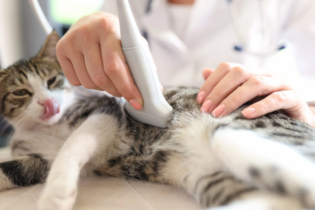
Imaging Should Be Part of the Initial Diagnostic Work-up
Diagnostic imaging remains the cornerstone of confirming feline urolithiasis. Survey abdominal radiography is generally the first-line imaging modality because it readily detects most radiopaque uroliths, including calcium oxalate calculi larger than approximately 1 mm². Besides confirming the presence of stones, radiographs help determine their number, size, location, shape and radiographic opacity, all of which influence therapeutic planning¹˒².
When performing radiography, a right lateral projection offers excellent visualisation of the urinary bladder, whereas the bladder neck may be partially obscured on ventrodorsal views².
Radiography, however, does not detect every urinary abnormality. Ultrasonography should complement radiographic evaluation, particularly when radiographs are inconclusive or radiolucent stones are suspected. Ultrasonographically, bladder uroliths typically appear as hyperechoic structures producing a distinct distal acoustic shadow and often move to the dependent portion of the bladder when the patient's position changes². Ultrasound also enables assessment of bladder wall thickness, intraluminal debris, blood clots and other concurrent abnormalities that may influence diagnosis¹˒².
Clinical Pearl: Radiography tells you whether stones are present; ultrasonography helps explain their clinical significance.
Urinalysis Supports the Diagnosis—but Does Not Confirm It
Urinalysis remains an essential part of the diagnostic work-up but should always be interpreted alongside imaging findings. The presence of urinary crystals suggests supersaturation of stone-forming minerals but does not confirm urolithiasis, while some cats with confirmed urinary stones may have no crystalluria at all¹.
Similarly, urine pH should be interpreted cautiously. Although acidic urine may favour calcium oxalate formation and alkaline urine may favour struvite crystallisation, urine pH alone cannot reliably predict stone composition¹.
Microscopic urine sediment examination can provide additional clues. Haematuria, leukocyturia, epithelial cells and proteinuria commonly reflect irritation and inflammation secondary to urolithiasis rather than primary renal disease². Prompt examination of fresh urine samples is essential because storage can alter crystal formation and sediment characteristics¹.
Don't Overlook Blood Biochemistry
Serum biochemistry provides valuable information, particularly in cats with suspected urinary obstruction. Measurement of blood urea nitrogen (BUN) and creatinine helps identify post-renal azotemia resulting from urethral obstruction or severe urinary outflow impairment². In addition, evaluating serum calcium is worthwhile, especially in cats with calcium oxalate urolithiasis, as underlying hypercalcaemia may contribute to stone formation and recurrence¹.
Take-Home Message
Successful diagnosis of feline urolithiasis depends on recognising that clinical signs alone are rarely diagnostic. A structured approach combining history, physical examination, urinalysis, serum biochemistry and, most importantly, early diagnostic imaging allows veterinarians to distinguish urolithiasis from other causes of FLUTD more confidently. Rather than viewing radiography, ultrasonography and laboratory testing as competing investigations, clinicians should consider them complementary tools that together improve diagnostic accuracy and facilitate timely, evidence-based treatment.
References (Vancouver)
- Bartges JW. Feline calcium oxalate urolithiasis: risk factors and rational treatment approaches. Journal of Feline medicine and Surgery. 2016 Sep;18(9):712-22. https://journals.sagepub.com/doi/full/10.1177/1098612x16660442
- Yaygıngül R. Clinical, laboratory, radiography and ultrasonography findings and surgical treatment the lower urinary system urolithiasis in cats and dogs. Animal Health Production and Hygiene. 2024 Jun 6;13(1):23-30. https://dergipark.org.tr/en/download/article-file/347265
Related Contents

Upcoming Event
ECG Interpretation Made Easy for Small Animal Practitioners
Electrocardiography (ECG) is an essential diagnostic tool in small animal practice, yet many clinici...

Upcoming Event
Positive Inotropes and Their Role in Cardiology
Positive inotropic agents are commonly used in cardiology to improve myocardial contractility and su...

Upcoming Event
Lesion-Based Diagnosis of Economically Important Poultry Diseases: A Visual Journey Through Gross Pathology
Accurate recognition of gross pathological lesions is essential for the diagnosis and control of pou...
.jpg)
Upcoming Event
Otitis in Cats and Dogs
Otitis is one of the most common ear disorders affecting the health and comfort of cats and dogs. Ga...

Upcoming Event
Postpartum Reproductive Disorders in Dairy Cattle
Postpartum reproductive disorders are a major cause of reduced fertility and economic losses in dair...
.jpg)
Upcoming Event
Bird Flu: A Bird's-Eye View
Avian influenza, commonly known as bird flu, remains one of the most significant infectious diseases...
.jpg)
Upcoming Event
Bovine Tuberculosis: Diagnostic Challenges and Pathological Features
Bovine tuberculosis is a chronic infectious disease that continues to impact cattle health, farm eco...

Upcoming Event
Post-Mortem Examination: Practical Tips for Field Veterinarians
Post-mortem examination is a valuable diagnostic tool that helps veterinarians determine the cause o...