Article
When Canine Atopic Dermatitis Treatment Fails: Finding the Missing Piece
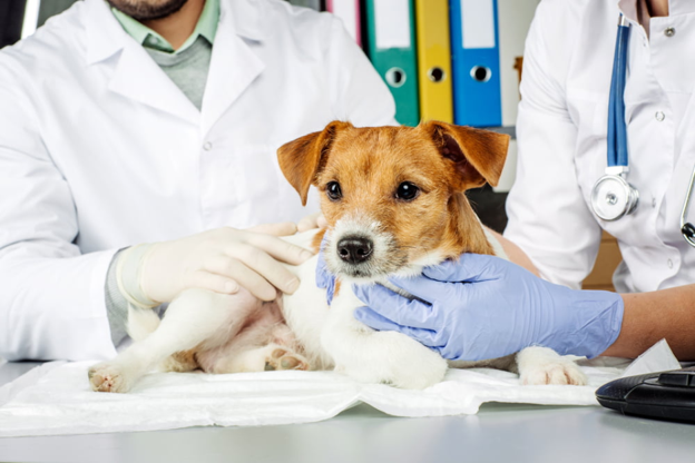
A six-year-old French Bulldog with a history of canine atopic dermatitis (CAD) returns to the clinic three months after starting oclacitinib. Initially, the itching reduced dramatically, and both the dog and the owner were finally getting some relief. But today, the story is different. The dog is scratching constantly, the ears are inflamed again, there is brown discharge between the toes, and the owner is convinced that "the medicine has stopped working.
Sound familiar?
For many veterinarians, these are among the most challenging dermatology consultations. However, true drug failure is often not the reason. More commonly, another factor has developed alongside CAD, turning a previously controlled patient into one with recurrent flare-ups. Before switching medications or increasing the dose, it is worth asking one simple question:
What has changed?
Treatment Failure—or Disease Flare?
Canine atopic dermatitis is a chronic, relapsing disease. Even well-controlled patients experience flare-ups when additional triggers overwhelm the skin's already compromised barrier1.
In many cases, persistent itching is not caused by inadequate immunomodulation but by factors that continue to drive inflammation despite ongoing therapy. Identifying these triggers is often the key to restoring disease control.
Secondary Infections: The Most Common Culprit
Secondary bacterial pyoderma and Malassezia dermatitis are among the leading causes of apparent treatment failure1.
Dogs may present with:
- Increased pruritus
- Erythematous paws
- Recurrent otitis
- Greasy skin
- Hyperpigmentation
- Lichenification
- New papules or pustules
These infections amplify inflammation, making owners believe that the primary medication is no longer effective.
Rather than immediately changing systemic therapy, repeat cytology should become routine whenever a CAD patient deteriorates. Identifying cocci, rods, or yeast allows targeted antimicrobial therapy that often restores clinical improvement without altering the underlying allergy treatment2.
Has the Skin Barrier Broken Down Again?
The skin barrier in dogs with CAD is inherently fragile. Once compromised, allergens, irritants, and microorganisms penetrate more easily, perpetuating inflammation and increasing susceptibility to infection1.
Barrier dysfunction can worsen because of:
- Excessive scratching
- Repeated infections
- Infrequent topical therapy
- Environmental allergens
- Seasonal changes
A dog whose itching appears poorly controlled may actually be suffering from progressive barrier damage rather than inadequate immunosuppression.
Regular bathing with therapeutic shampoos, moisturising products, and barrier-supportive topical therapies can significantly improve long-term skin health.
Don't Forget the Other Allergies
Not every flare is caused solely by environmental atopy.
Concurrent flea allergy dermatitis remains an important differential diagnosis, particularly in regions where flea exposure is common. Likewise, adverse food reactions can coexist with environmental allergies, making clinical signs more severe and more difficult to control1.
If flare-ups persist despite apparently appropriate treatment, reviewing parasite prevention and considering a dietary elimination trial may be worthwhile before modifying long-term medication.
Could Owner Compliance Be the Real Problem?
Managing CAD is demanding.
Dogs often require long-term medication, topical therapy, specialised diets, parasite control, and regular rechecks. Over time, treatment fatigue may develop, leading to skipped doses, irregular bathing, or inconsistent diet adherence.
Rather than assuming pharmacological failure, discussing the owner's daily management routine often reveals practical barriers that can be corrected through education and simplified treatment plans.
Use Cytology Before You Change Drugs
One of the most valuable diagnostic tools in dermatology remains skin cytology.
Whenever a patient experiences recurrent itching despite therapy, cytology can quickly identify bacterial infection or yeast overgrowth and guide appropriate treatment.
Escalating immunomodulatory therapy without first investigating secondary infections may prolong disease, increase antimicrobial use later, and leave the underlying cause unresolved.
A Practical Checklist for the Non-Responsive CAD Patient
Before concluding that treatment has failed, ask yourself:
✔ Has the diagnosis of CAD been confirmed?
✔ Has repeat cytology been performed?
✔ Is there bacterial pyoderma or Malassezia overgrowth?
✔ Is parasite control up to date?
✔ Could a concurrent food allergy be contributing?
✔ Is the owner following the treatment plan consistently?
✔ Does the skin barrier require additional support?
Working systematically through these questions often identifies the missing piece responsible for persistent disease.
The Clinical Take-Home
When a dog with canine atopic dermatitis stops responding to treatment, resist the temptation to simply reach for a different medication. In many cases, the underlying therapy is still working—but secondary infections, skin barrier damage, concurrent allergies, or management issues have become the new drivers of disease. A structured reassessment that includes repeat cytology, evaluation of barrier health, parasite control, and owner compliance frequently restores disease control without escalating immunosuppressive therapy. For difficult CAD cases, the best next treatment is often a better investigation rather than a stronger drug.
References (Vancouver)
- Santoro D, Marsella R, Pucheu-Haston CM, Eisenschenk MNC, Nuttall T, Bizikova P. Current knowledge on canine atopic dermatitis: pathogenesis, diagnosis and treatment. Vet Sci. 2022;9(7):330. doi:10.3390/vetsci9070330. Available from: https://www.mdpi.com/2306-7381/9/7/330
- Olivry T, DeBoer DJ, Favrot C, Jackson HA, Mueller RS, Nuttall T, et al. Treatment of canine atopic dermatitis: updated guidelines from the International Committee on Allergic Diseases of Animals (ICADA). BMC Vet Res, 2015. Available from: https://doi.org/10.1186/s12917-015-0514-6
Related Contents

Upcoming Event
ECG Interpretation Made Easy for Small Animal Practitioners
Electrocardiography (ECG) is an essential diagnostic tool in small animal practice, yet many clinici...

Upcoming Event
Positive Inotropes and Their Role in Cardiology
Positive inotropic agents are commonly used in cardiology to improve myocardial contractility and su...

Upcoming Event
Lesion-Based Diagnosis of Economically Important Poultry Diseases: A Visual Journey Through Gross Pathology
Accurate recognition of gross pathological lesions is essential for the diagnosis and control of pou...
.jpg)
Upcoming Event
Otitis in Cats and Dogs
Otitis is one of the most common ear disorders affecting the health and comfort of cats and dogs. Ga...

Upcoming Event
Postpartum Reproductive Disorders in Dairy Cattle
Postpartum reproductive disorders are a major cause of reduced fertility and economic losses in dair...
.jpg)
Upcoming Event
Bird Flu: A Bird's-Eye View
Avian influenza, commonly known as bird flu, remains one of the most significant infectious diseases...
.jpg)
Upcoming Event
Bovine Tuberculosis: Diagnostic Challenges and Pathological Features
Bovine tuberculosis is a chronic infectious disease that continues to impact cattle health, farm eco...

Upcoming Event
Post-Mortem Examination: Practical Tips for Field Veterinarians
Post-mortem examination is a valuable diagnostic tool that helps veterinarians determine the cause o...