Article
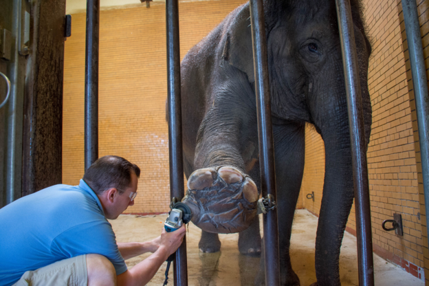
Elephant Foot Health: Detecting Problems Before Lameness
Foot problems in elephants do not necessarily begin with obvious lameness. By the time an elephant is visibly reluctant to bear weight or its gait has clearly changed, the underlying problem may already have progressed.
This makes routine foot assessment more than a search for open wounds or overgrown nails. The objective is to recognise small, persistent or progressive changes that may warrant closer evaluation before locomotor impairment becomes apparent.
Start with what can be seen—and document it
A structured examination should assess the sole, nails, interdigital spaces and surrounding tissues for changes in shape, surface, swelling, cracks, discharge, abnormal tissue growth or asymmetry.
The value of the examination increases when findings are documented consistently. Serial photographs and records can help establish whether a defect is stable, improving or gradually changing—something that a single examination may not reveal.
This is particularly useful for elephants undergoing regular foot care, because the current appearance can be compared with the animal's own previous findings rather than relying only on a general description of what is considered normal.
A small external lesion may not explain the whole problem
The external appearance of the foot does not always indicate what is happening deeper within it.
Computed tomography of 52 elephant feet identified skeletal abnormalities including bone remodelling, enthesopathy, osteoarthritis, osseous cyst-like lesions, osteitis and infectious osteoarthritis. Importantly, several feet showed more than one type of pathological change, demonstrating that abnormalities may coexist within the same foot[1].
For clinical assessment, this means that a seemingly minor external lesion should not automatically be assumed to account for persistent discomfort, swelling or locomotor change.
If the clinical picture appears disproportionate to the visible lesion—or if a problem repeatedly returns—the possibility of deeper pathology deserves consideration.
Imaging findings need clinical correlation
Advanced imaging can reveal abnormalities that cannot be appreciated externally, but an unusual appearance on imaging is not automatically disease.
The same CT study documented substantial anatomical variation in elephant phalanges and sesamoid bones[1]. Some unusual structures could therefore be mistaken for pathology if imaging findings are interpreted without considering normal anatomical variation.
The relevant question is not simply:
“Is this structure abnormal?”
It is:
“Does this finding fit the clinical signs, anatomical location and overall pattern of disease in this elephant?”
That distinction is particularly important when deciding whether an imaging finding is clinically meaningful.
Recurrent foot problems warrant a wider review
When a foot lesion repeatedly recurs, examining the foot alone may not be enough.
Management and environmental conditions should form part of the clinical history. In a study of African and Asian elephants housed in North American zoos, foot and musculoskeletal health were associated with factors including time spent on hard substrates, available space and indoor/outdoor housing patterns[2]. Because the study was observational and conducted in North American zoos, these associations should not be interpreted as evidence that a particular substrate directly causes foot disease in Indian elephants. However, they support considering housing, substrate and activity when evaluating recurrent foot or musculoskeletal problems[2].
For elephants managed in India, this makes the environmental history relevant when a lesion persists despite appropriate local foot care.
When infection is a concern, characterise the lesion
A chronic, non-healing or clinically infected foot lesion may require investigation beyond visual examination.
In a 2024 study of captive Asian elephants in India, bacterial organisms were isolated from samples collected from foot lesions, with both Gram-negative and Gram-positive bacteria identified[3]. Escherichia coli, Bacillus spp., Pseudomonas spp. and Staphylococcus spp. were among the organisms reported[3].
These findings should not be interpreted as proof that every isolated organism was the primary cause of the lesion. The study identified bacterial flora associated with lesions; it did not establish pathogenicity for every isolate.
The practical implication is more measured: when infection is clinically suspected, appropriate sampling can provide information that visual assessment alone cannot.
When should further investigation be considered?
Further investigation becomes more relevant when:
- A lesion persists or repeatedly recurs;
- Swelling cannot be explained by the external findings;
- The elephant develops persistent or progressive lameness;
- The severity of clinical signs appears disproportionate to the visible lesion;
- Infection is suspected;
- The response to management is poorer than expected.
The next diagnostic step should be determined by the clinical question. Where deeper pathology is suspected, imaging may help identify changes that are not externally visible. Where infection is suspected, appropriate microbiological investigation may provide additional information.
The practical point
The aim of preventive foot assessment is not simply to find disease—it is to recognise change early enough to investigate it appropriately.
A consistent examination, longitudinal documentation and attention to environmental factors can help identify problems before obvious lameness develops. When findings do not fit the clinical picture, deeper pathology or infection should remain considerations rather than assuming that the visible lesion tells the entire story.
Reference(s):
- Regnault S, Dixon JJ, Warren-Smith C, Hutchinson JR, Weller R. Skeletal pathology and variable anatomy in elephant feet assessed using computed tomography. PeerJ. 2017;5:e2877. Published 2017 Jan 18. https://doi.org/10.7717/peerj.2877
- Miller MA, Hogan JN, Meehan CL. Housing and demographic risk factors impacting foot and musculoskeletal health in African elephants [Loxodonta africana] and Asian elephants [Elephas maximus] in North American zoos. PLoS One. 2016 Jul 14;11(7):e0155223. https://doi.org/10.1371/journal.pone.0155223
- Veeraselvam M, Selvaraj P, Kumar SS, Senthilkumar A, Senthilkumar T, Jayalakshmi K, et al. Isolation and Identification of Bacterial Flora from Foot Lesions of Asian Elephants (Elephas maximus). UPJOZ [Internet]. 2024 Mar. 14 [cited 2026 Sep. 1];45(6):22-8. https://mbimph.com/index.php/UPJOZ/article/view/3948
Related Contents
Article
When an Elephant’s Behaviour Changes: Recognising Clinically Relevant Patterns
A change in an elephant’s behaviour can be easy to dismiss as temperament. It can also...
Article
Is It Normal for This Elephant? Building an Individual Health Baseline
For a long-lived animal, a single health assessment provides only a snapshot. A more useful c...
Article
Body Condition in Asian Elephants: When Does Change Become Clinically Relevant?
Body condition is often recorded during routine elephant health assessments. Its clinical value, how...
Article
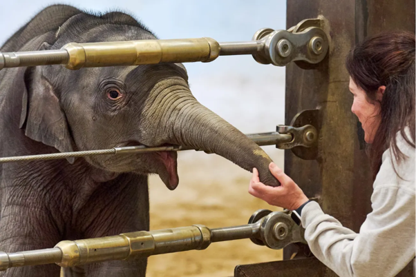
EEHV in Asian Elephant Calves: Recognising the Early Clinical Picture
Elephant endotheliotropic herpesvirus (EEHV) is an important cause of fatal haemorrhagic disease in...
Article
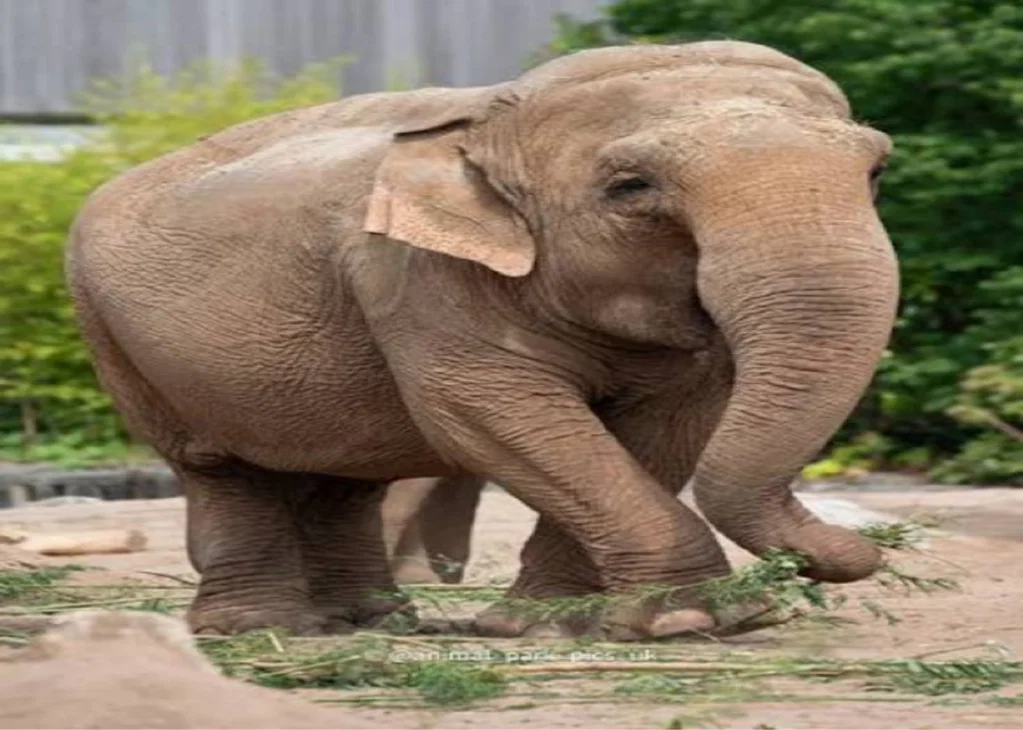
Gastrointestinal Parasites in Asian Elephants: Interpreting Faecal Findings
A positive faecal examination confirms the detection of parasite stages in the sample. It does not,...
Article
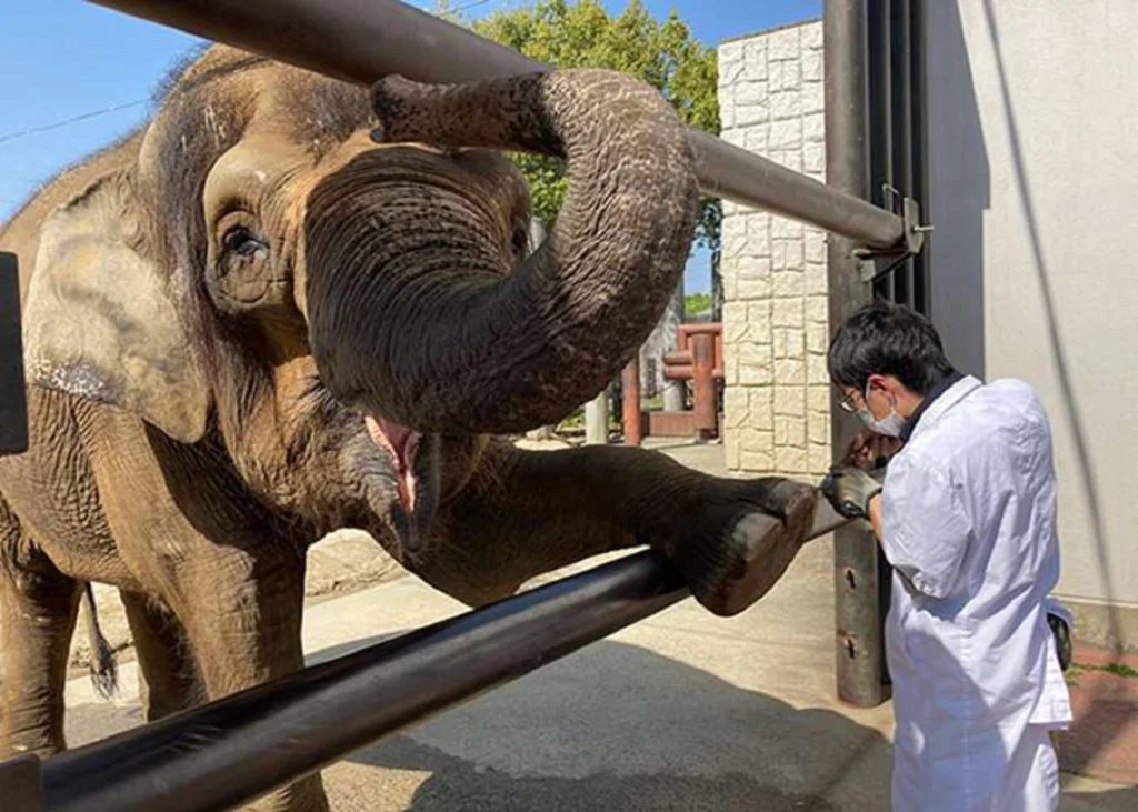
Tuberculosis in Asian Elephants: Interpreting Screening Results in Context
Tuberculosis surveillance in elephants presents a fundamental diagnostic challenge: a test result is...
Article
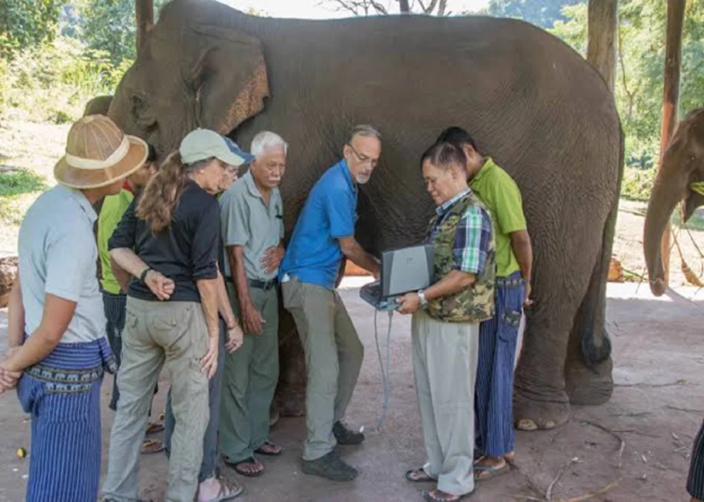
Pregnancy Monitoring in Asian Elephants: Interpreting Ultrasound and Hormonal Findings
Confirming pregnancy is only the beginning of reproductive monitoring in an elephant. With a...
Article
When an Elephant Stops Eating: Interpreting Changes in Appetite and Feeding Behaviour
A reduction in food intake can be one of the earliest observable changes in an elephant that is beco...