Article
Concurrent Chorioptes Infestation and Dermatophytosis in Goats: A Practical Diagnostic Approach
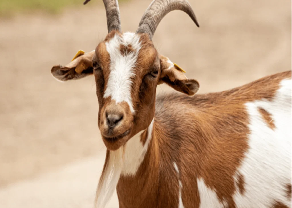
Crusting, scaling and alopecia in a goat can point towards an ectoparasitic problem, but the severity and appearance of the lesions may tell a different story. Chorioptes bovis infestation is particularly common in goats and is often subclinical or asymptomatic. When skin lesions become more extensive or clinically severe, concurrent dermatophytosis should also be considered.
When Chorioptes is Not the Whole Picture
Chorioptes bovis primarily affect the feet, where it can cause crust formation or “foot mange”. Small crusts may also remain concealed beneath the hair coat. Because the mites feed on skin debris rather than causing extensive tissue damage, infection in goats is often subclinical[1].
This makes the clinical presentation important. Marked alopecia, scaling and crusting may resemble dermatophytosis, and the presence of another skin infection can intensify the clinical picture[1],[2].
Look Beyond the Visible Lesion[2]
Dermatophytosis in goats is less frequently reported than in cattle, but Trichophyton verrucosum and T. mentagrophytes are among the dermatophytes reported in this species.
Direct examination of skin samples is particularly useful when crusts and scales are present. Thick keratin-rich material can be cleared using NaOH or KOH, allowing fungal hyphae and conidia to be visualised. In affected goats, this approach can also help identify the organisms responsible for the dermatological lesions.
For T. verrucosum, infected hairs within crusts and scales may provide useful diagnostic material. Large arthroconidia arranged in chains or groups can be suggestive of this dermatophyte and help distinguish it from other dermatophytes affecting livestock.
Culture Has a Role Too[2]
Direct microscopy provides rapid information, while culture can establish fungal identity through morphological and molecular assessment.
One practical limitation is the slow growth of T. verrucosum. Cultures may require several days to become positive, while faster-growing non-pathogenic moulds can contaminate animal hair and complicate isolation.
Keep the Herd and People in Mind[2]
The diagnostic distinction is also important from a transmission perspective. Dermatophytosis is contagious, and T. verrucosum can spread to humans through infected animals, contaminated fomites, or soil.
For herd-level management, apparently unaffected goats may also warrant attention when dermatophytosis and chorioptic infestation occur together. In the reported case, asymptomatic animals were included in treatment because subclinical lesions could not be completely excluded, and untreated animals could potentially allow continued spread.
Clinical Bottom Line
A goat with crusting and alopecia may have more than one dermatological problem. Chorioptes infestation should be assessed alongside dermatophytosis when lesions are unusually severe or widespread. Direct examination of appropriately collected crusts, scales and hairs can provide rapid diagnostic direction, while culture can support definitive fungal identification.
The combination of clinical assessment, direct microscopy and appropriate herd-level attention can help avoid attributing the entire presentation to a mite infestation alone.
References:
- Rambozzi L, Meneguz PG, Molinar Min AR, Pasquetti M, Peano A. Concurrent Chorioptic mange and Dermatophytosis in dairy goats: A case report. Veterinary Sciences. 2022 Dec 6;9(12):677. https://www.mdpi.com/2306-7381/9/12/677
- Hameed K, Ch FR, Nawaz MA, Naqvi SM, Gräser Y, Kupsch C, Pasquetti M, Rossi L, Min AR, Tizzani P, Chiavassa E. Trichophyton verrucosum infection in livestock in the Chitral district of Pakistan. The journal of infection in developing countries. 2017 Apr 30;11(04):326-33. https://jidc.org/index.php/journal/article/download/28459224/1689
Related Contents
Article
Sudden Death in Cattle: A Practical Differential Diagnosis
A cow is found dead during the morning inspection. There were no obvious warning signs the previous...
Article
Caudal Vena Cava Thrombosis in Cattle: Recognising a Fatal Complication of Liver Abscessation
A cow may appear completely healthy until sudden respiratory distress, haemoptysis or death occurs....
Article
Sudden Death in Sheep: Key Differential Diagnoses for the Veterinarian
A sheep found dead on pasture may leave little warning. One animal may have been affected by a rapid...
Article
Sudden Death in Goats: Recognising Peracute Infectious and Metabolic Causes
A goat found dead with little or no warning can leave very few clinical clues. In these cases, the p...
Article
Necrotic Enteritis in Broilers: Identifying the Farm-Level Risk Factors
A broiler flock showing wet litter, poor performance or rising mortality may have several problems o...
Article
Necrotic Enteritis in Broilers: Linking Intestinal Lesions with Laboratory Diagnosis
A broiler may arrive for postmortem examination with a distended, fragile small intestine covered by...
Article
Controlling Necrotic Enteritis Without Antibiotic Growth Promoters: What Can Feed-Based Strategies Offer?
When antibiotic growth promoters are no longer the main option for controlling necrotic enteritis (N...
Article
Necrotic Enteritis and Antimicrobial Resistance: Implications for Poultry and Food Safety
When a broiler flock develops necrotic enteritis (NE), antimicrobial treatment may appear to be the...