Article
Neonatal Calf Diarrhea: Understanding the Major Infectious Causes
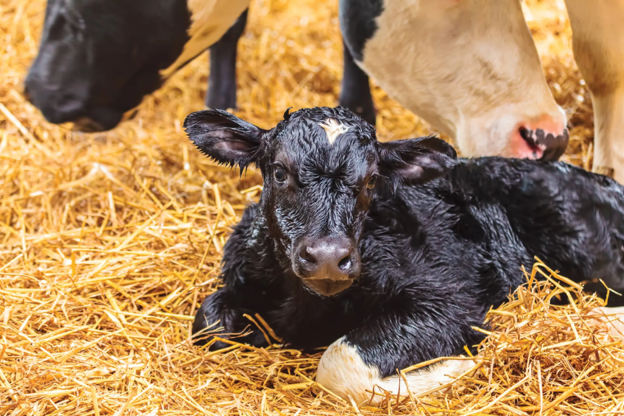
Neonatal calf diarrhea remains one of the most significant health challenges encountered in calf-rearing operations. Beyond its impact on calf welfare, diarrhea contributes substantially to mortality, morbidity, treatment costs, and reduced productivity. Although diarrhea is often discussed as a single disease entity, practicing veterinarians know that it is usually a multifactorial condition involving complex interactions between pathogens, host factors, and environmental conditions.
Several infectious agents are commonly associated with neonatal calf diarrhea, including bacteria, viruses, and protozoa. Importantly, many of these organisms can also be detected in healthy calves, meaning their presence alone does not always confirm disease. Understanding the pathophysiology of the major enteric pathogens can help guide clinical assessment and management decisions1.
Bacterial Causes of Diarrhea
Among bacterial pathogens, enterotoxigenic Escherichia coli (ETEC) is considered the most prevalent cause of neonatal calf diarrhea, particularly in calves younger than four days of age2.
ETEC attaches to intestinal epithelial cells through fimbrial antigens, with the K99 antigen being particularly important1. Once attached, heat-stable toxins stimulate chloride secretion into the intestinal lumen. Water follows this electrolyte movement, resulting in secretory diarrhea1,2. Because the intestinal epithelium remains largely intact, ETEC-associated diarrhea is typically non-hemorrhagic.
Other E. coli pathogroups, including enteropathogenic E. coli (EPEC), Shiga toxin-producing E. coli (STEC), attaching and effacing E. coli (AEEC), and enterohaemorrhagic E. coli (EHEC), may contribute to diarrhea in older calves2. Unlike ETEC, these organisms can damage the intestinal epithelium and may be associated with hemorrhagic diarrhea.
Salmonella spp., particularly S. typhimurium, are also important enteric pathogens in calves2. These organisms invade intestinal epithelial cells and stimulate inflammatory cytokine release, leading to inflammation, ulceration, and mucosal damage1. The resulting impairment of water and electrolyte absorption contributes to clinical diarrhea and systemic illness.
The role of Clostridium perfringens remains less clear. Although toxin-mediated intestinal damage and fluid loss have been described, the organism is also a normal inhabitant of the calf gastrointestinal tract, making interpretation of diagnostic findings challenging1.
Viral Enteropathogens
Bovine coronavirus (BCoV) and bovine rotavirus (BRoV) are among the most important viral causes of neonatal calf diarrhea2.
BCoV affects both the small and large intestines. Infection leads to destruction of intestinal villi and replacement of mature epithelial cells with immature cells that have reduced absorptive and digestive capacity3. The result is malabsorptive and hyperosmotic diarrhea.
BRoV primarily targets mature enterocytes located on the villi of the small intestine1,4. Damage to these cells reduces nutrient and fluid absorption. In addition, the viral enterotoxin NSP4 disrupts normal fluid movement across the intestinal epithelium, promoting hypersecretory diarrhea1,2.
Cryptosporidium: A Major Protozoal Challenge
Cryptosporidium parvum is one of the most important causes of diarrhea in neonatal calves worldwide. Clinical disease is most commonly observed during the first few weeks of life, particularly in calves younger than 20 days5.
After ingestion, sporozoites invade the intestinal epithelium and establish infection5. Damage to villi reduces the absorptive surface area of the intestine, leading to fluid malabsorption and diarrhea. Increased intestinal permeability and prostaglandin-mediated fluid secretion may further contribute to disease severity1,5.
Clinical presentations can vary considerably. Some calves develop mild self-limiting diarrhea, while others experience severe disease, including hemorrhagic diarrhea1.
Practical Clinical Insights
For practicing veterinarians, neonatal calf diarrhea should rarely be viewed as a single-pathogen disease. Multiple infectious agents may be present simultaneously, and pathogen detection must always be interpreted alongside clinical findings, age of the calf, herd history, and disease patterns.
Recognizing age-associated pathogen distribution can be particularly useful during field investigations. ETEC tends to predominate in very young calves, whereas rotavirus, coronavirus, Salmonella spp., and C. parvum often become more relevant as calves age.
Key Takeaway
Successful management of neonatal calf diarrhea begins with understanding the distinct mechanisms through which bacterial, viral, and protozoal pathogens disrupt intestinal function. Identifying likely pathogens based on calf age, clinical presentation, and herd context can support more informed diagnostic and management decisions.
References
- Jessop E, Li L, Renaud DL, Verbrugghe A, Macnicol J, Gamsjäger L, Gomez DE. Neonatal calf diarrhea and gastrointestinal microbiota: etiologic agents and microbiota manipulation for treatment and prevention of diarrhea. Veterinary Sciences. 2024 Feb 29;11(3):108. https://www.mdpi.com/2306-7381/11/3/108
- Cho YI, Yoon KJ. An overview of calf diarrhea-infectious etiology, diagnosis, and intervention. Journal of veterinary science. 2014 Mar 19;15(1):1. https://synapse.koreamed.org/pdf/10.4142/jvs.2014.15.1.1
- Vlasova AN, Saif LJ. Bovine coronavirus and the associated diseases. Frontiers in Veterinary Science. 2021 Mar 31;8:643220. https://www.frontiersin.org/journals/veterinary-science/articles/10.3389/fvets.2021.643220/pdf
- Geletu US, Usmael MA, Bari FD. Rotavirus in calves and its zoonotic importance. Veterinary Medicine International. 2021;2021(1):6639701. https://onlinelibrary.wiley.com/doi/pdf/10.1155/2021/6639701
- Thomson S, Hamilton CA, Hope JC, Katzer F, Mabbott NA, Morrison LJ, Innes EA. Bovine cryptosporidiosis: impact, host-parasite interaction and control strategies. Veterinary research. 2017 Aug 11;48(1):42. https://link.springer.com/content/pdf/10.1186/s13567-017-0447-0.pdf
Related Contents

Upcoming Event
ECG Interpretation Made Easy for Small Animal Practitioners
Electrocardiography (ECG) is an essential diagnostic tool in small animal practice, yet many clinici...

Upcoming Event
Positive Inotropes and Their Role in Cardiology
Positive inotropic agents are commonly used in cardiology to improve myocardial contractility and su...

Upcoming Event
Lesion-Based Diagnosis of Economically Important Poultry Diseases: A Visual Journey Through Gross Pathology
Accurate recognition of gross pathological lesions is essential for the diagnosis and control of pou...
Upcoming Event
Postpartum Reproductive Disorders in Dairy Cattle
Postpartum reproductive disorders are a major cause of reduced fertility and economic losses in dair...
.jpg)
Upcoming Event
Bird Flu: A Bird's-Eye View
Avian influenza, commonly known as bird flu, remains one of the most significant infectious diseases...
.jpg)
Upcoming Event
Bovine Tuberculosis: Diagnostic Challenges and Pathological Features
Bovine tuberculosis is a chronic infectious disease that continues to impact cattle health, farm eco...

Upcoming Event
Post-Mortem Examination: Practical Tips for Field Veterinarians
Post-mortem examination is a valuable diagnostic tool that helps veterinarians determine the cause o...

Upcoming Event
PCR, Serology, or Culture? Choosing the Right Diagnostic Test for Zoonotic Diseases
Confused between PCR, serology, and culture for diagnosing zoonotic diseases? Learn how to se...