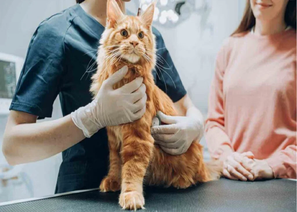
Article
Can Renal Fibrosis Biomarkers Reveal CKD Progression in Cats?
Chronic kidney disease in cats is not only a problem of declining renal function. Structural changes within the kidney, including fibrosis, can also develop as chronic renal injury progresses. This makes biomarkers associated with fibrotic activity potentially useful for identifying changes that may not be fully reflected by routine renal function markers.
Two biomarkers investigated in feline CKD are transforming growth factor-beta 1 (TGF-β1) and procollagen type III N-terminal propeptide (PIIINP). Both provide information related to renal fibrosis, but their clinical interpretation requires caution.
TGF-β1: Looking for Fibrotic Activity
TGF-β1 is involved in the development of renal fibrosis and has therefore been investigated as a urinary biomarker in cats with CKD. Studies have reported higher urinary total TGF-β1 concentrations in cats with CKD compared with healthy cats[1],[2].
However, the relationship between urinary TGF-β1 and renal fibrosis is not straightforward. Differences in assays and the forms of TGF-β1 being measured can affect the results. One investigation evaluated the relationship between urinary TGF-β1 and renal fibrosis and highlighted the need for appropriate assay interpretation[3].
An interesting finding was that the urinary active TGF-β1-to-creatinine ratio (uaTGF-β1/Cr) increased approximately 2.6-fold before cats subsequently developed azotemia[3]. This raises the possibility that TGF-β1 may provide information about ongoing renal changes before conventional evidence of azotemia becomes apparent.
However, an elevated TGF-β1 concentration should not automatically be interpreted as a direct measurement of the amount of fibrosis present. The biomarker reflects a biological process associated with fibrosis rather than providing a direct assessment of renal structural change.
PIIINP: A Marker Related to Collagen Formation
Procollagen type III N-terminal propeptide (PIIINP) is associated with type III collagen formation and has been investigated as another potential marker of renal fibrosis. In cats with azotemic CKD, urinary PIIINP-to-creatinine ratio (uPIIINP/Cr) was increased and showed a high correlation with renal parenchyma stiffness measured by shear-wave ultrasonography[4].
This finding is relevant because it connects a urinary biomarker with a measurable change in renal tissue characteristics. However, feline assay validation was not reported, which limits how confidently uPIIINP/Cr can currently be applied as a routine clinical biomarker.
What Should You Take From TGF-β1 and PIIINP?[2]
TGF-β1 and PIIINP can add information about what is happening within a chronically injured kidney, rather than only showing how well the kidneys are filtering.
For TGF-β1, an increase in urinary active TGF-β1 has been reported before some cats subsequently developed azotemia. This makes it particularly interesting when looking for signs of ongoing renal change before azotemia becomes evident.
PIIINP provides a different clue. Increased urinary PIIINP-to-creatinine ratios in azotemic CKD cats were associated with greater renal parenchyma stiffness, linking the biomarker with changes in renal tissue.
However, neither marker should be used alone to determine the severity of renal fibrosis. TGF-β1 results can vary depending on the assay and the form measured, while feline data on PIIINP remains limited.
The practical point: these biomarkers may offer additional information about renal fibrotic activity, but they need to be interpreted alongside the cat’s overall CKD findings rather than as standalone measures of fibrosis.
References:
- Habenicht LM, Webb TL, Clauss LA, Dow SW, Quimby JM. Urinary cytokine levels in apparently healthy cats and cats with chronic kidney disease. Journal of feline medicine and surgery. 2013 Feb;15(2):99-104. https://journals.sagepub.com/doi/pdf/10.1177/1098612X12461007
- Kongtasai T, Paepe D, Meyer E, Mortier F, Marynissen S, Stammeleer L, Defauw P, Daminet S. Renal biomarkers in cats: A review of the current status in chronic kidney disease. Journal of veterinary internal medicine. 2022 Mar;36(2):379-96. https://academic.oup.com/jvim/article-pdf/36/2/379/66666729/jvim16377.pdf
- Lawson JS, Syme HM, Wheeler-Jones CP, Elliott J. Urinary active transforming growth factor β in feline chronic kidney disease. The Veterinary Journal. 2016 Aug 1;214:1-6. https://www.sciencedirect.com/science/article/pii/S1090023316000356
- Thanaboonnipat C, Sutayatram S, Buranakarl C, Choisunirachon N. Renal shear wave elastography and urinary procollagen type III amino-terminal propeptide (uPIIINP) in feline chronic kidney disease. BMC veterinary research. 2019 Feb 11;15(1):54. https://link.springer.com/content/pdf/10.1186/s12917-019-1801-4.pdf
Related Contents
Article
FGF-23 in Feline CKD: An Early Signal of Phosphate Dysregulation
A cat with early chronic kidney disease (CKD) may have a normal serum phosphate concentration. Does...
Article
Albuminuria or Transferrinuria? Reading the Glomerular Signal in Feline CKD
A cat can have chronic kidney disease (CKD) with only mild proteinuria. So, when urinary protein is...
Article
Tubular Injury Biomarkers in Feline CKD: What Can NGAL, KIM-1 and L-FABP Tell You?
When feline CKD is already suspected, identifying reduced renal function is only part of the clinica...
Article
When Creatinine May Not Tell the Full Story in Feline Acute Kidney Injury
Serum creatinine is routinely used with changes in urine output to diagnose and grade Acute Kidney I...
Article
When a Normal-Looking Udder Hides Mastitis: Recognising Subclinical Mastitis in Buffaloes
A buffalo can appear clinically normal while mastitis is already affecting the udder and milk produc...
Article
Buffalo Subclinical Mastitis: Which Risk Factors Should Vets Investigate?
When subclinical mastitis (SCM) keeps appearing in a buffalo herd, looking only at the affected anim...

Article
Chronic Cough in Dogs and Cats: How to Classify and Localize It
A dog has been coughing for several weeks. The owner describes it as “honking,” but the...
Article
Diagnosing the Coughing Dog or Cat: Choosing the Right Diagnostic Test
A coughing dog or cat does not automatically need every available respiratory test. The more useful...