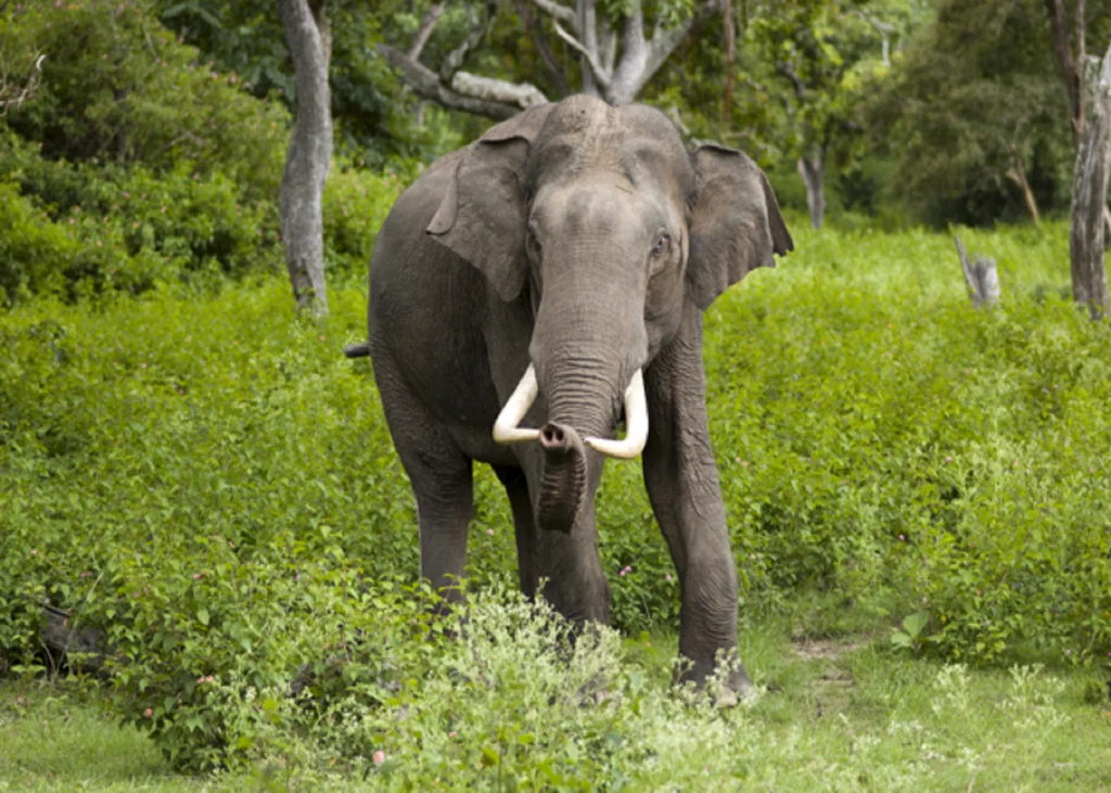
Article
Elephant Necropsy: Turning Post-Mortem Findings into a Better Health Investigation
An elephant necropsy should answer more than “What lesion was present?”
The greater diagnostic value comes from connecting the clinical history, gross findings, histopathology and targeted laboratory testing to determine what most likely contributed to death.
This is particularly important in elephants because mortality may be an uncommon event for an individual veterinary team, while the opportunity to investigate the carcass is unlikely to recur.
Start before the necropsy begins
The post-mortem investigation should begin with the case history.
Relevant information includes:
- Age and sex;
- Presenting clinical signs;
- Duration of illness;
- Laboratory findings;
- Imaging findings;
- Medications administered;
- Previous disease;
- Dietary and management history;
- Contact history;
- Circumstances surrounding death.
The recently published elephant necropsy protocol specifically recommends obtaining the clinical history and herd information before the examination, including known or suspected tuberculosis status[1].
This information helps the pathologist decide which findings require particular attention and which additional samples may be necessary.
Gross examination should be systematic
The size and anatomy of an elephant create practical challenges that are not encountered in routine domestic-animal necropsies.
The 2026 pictorial necropsy guide provides a structured approach covering external examination, dissection of the major organ systems, organ-specific examination and tissue sampling[1].
The value of such a systematic approach is not simply procedural.
It reduces the likelihood that diagnostically important tissues will be overlooked because the examination is focused only on the organ system suspected from the clinical history.
A gross lesion may need laboratory confirmation
A striking lesion can strongly direct the diagnostic investigation, but morphology alone may not establish the cause.
This is well illustrated by tuberculosis investigations in Indian Asian elephants.
In one free-ranging elephant from Karnataka, post-mortem examination identified disseminated pulmonary nodules with central caseation, enlarged lymph nodes and acid-fast organisms. PCR and sequencing subsequently confirmed Mycobacterium tuberculosis[2].
Similarly, investigations of wild Asian elephants in southern India identified granulomatous lesions and acid-fast bacilli, with molecular testing confirming M. tuberculosis in affected animals[3].
The practical implication is important:
When the gross or histological findings suggest an infectious disease, appropriate confirmatory samples should be collected before the carcass is disposed of.
Sample strategically, not indiscriminately
The necropsy protocol recommends collecting tissue suitable for different downstream investigations, including histology, microbiology and molecular testing[1].
The choice of sample should reflect the differential diagnosis.
Depending on the case, this may mean retaining appropriate portions of:
- Affected organs;
- Regional lymph nodes;
- Gastrointestinal tissues;
- Respiratory tissues;
- Liver and spleen;
- Kidney;
- Brain;
- Reproductive organs;
- Other visibly abnormal tissues.
The objective is not to collect every tissue without a plan.
It is to ensure that the suspected disease process can still be investigated after the gross examination has ended.
Infectious disease changes the safety requirements
An elephant necropsy can present both biological and physical hazards.
The 2026 protocol specifically addresses workplace safety, personal protective equipment, transmissible disease precautions, site preparation, equipment and carcass handling[1].
This is especially relevant when diseases with zoonotic potential are included in the differential diagnosis.
Tuberculosis is a clear example in the Indian context: M. tuberculosis has been confirmed in free-ranging Asian elephants in southern India, making appropriate precautions important when compatible lesions or epidemiological concerns are present[2]˒[3].
Do not separate pathology from epidemiology
The significance of a post-mortem finding may extend beyond the individual elephant.
For example:
- An infectious diagnosis may justify investigation of other elephants;
- A toxicological finding may prompt examination of feed or environmental exposure;
- A traumatic lesion may indicate a management or enclosure risk;
- Severe parasitism may support broader herd surveillance;
- Recurrent lesions may indicate a population-level problem.
A survey of veterinarians working with Asian elephants across eight range countries reported trauma, gastrointestinal disease, parasitism and infectious disease among important causes of morbidity and mortality, while also identifying lung lesions, gastrointestinal stasis or torsion, parasites and other pathological findings among reported necropsy observations[4].
The survey was based on veterinary respondents and therefore should not be interpreted as a population-level mortality database.
Its practical value is instead in illustrating the breadth of pathology that may be encountered and the need for better diagnostic capacity[4].
Document negative findings as carefully as positive ones
A good necropsy report should record what was examined as well as what was abnormal.
This creates a useful baseline if:
- Another elephant subsequently dies;
- A similar clinical syndrome occurs;
- Additional laboratory results become available;
- The case is reviewed retrospectively.
Photographs, measurements and properly labelled tissue samples can substantially increase the future diagnostic value of the examination.
The final diagnosis should reflect the strength of the evidence
Not every case will produce a definitive cause of death.
Where evidence is incomplete, the report should distinguish between:
- Confirmed diagnosis;
- Probable diagnosis;
- Suspected contributing condition;
- Incidental finding;
- Undetermined cause.
This is preferable to assigning a definitive cause when the available evidence does not support one.
Clinical takeaway
An elephant necropsy is most valuable when it is planned as a diagnostic investigation rather than treated as a final visual examination.
A systematic gross examination, appropriate tissue sampling, targeted laboratory testing and careful documentation can turn a single mortality event into useful information for the remaining population.
References:
- Falkenau A, Kolb N, Rieger A, et al. A pictural guide to postmortem examination of elephants. PLoS One. 2026;21(2):e0338783. Published 2026 Feb 9. https://doi.org/10.1371/journal.pone.0338783
- Chandranaik BM, Shivashankar BP, Umashankar KS, et al. Mycobacterium tuberculosis Infection in Free-Roaming Wild Asian Elephant. Emerg Infect Dis. 2017;23(3):555-557. https://doi.org/10.3201/eid2303.161439
- Zachariah A, Pandiyan J, Madhavilatha GK, et al. Mycobacterium tuberculosis in Wild Asian Elephants, Southern India. Emerg Infect Dis. 2017;23(3):504-506. https://doi.org/10.3201/eid2303.161741
- Miller D, Jackson B, Riddle HS, Stremme C, Schmitt D, Miller T. Elephant (Elephas maximus) Health and Management in Asia: Variations in Veterinary Perspectives. Vet Med Int. 2015;2015:614690. https://doi.org/10.1155/2015/614690
Related Contents
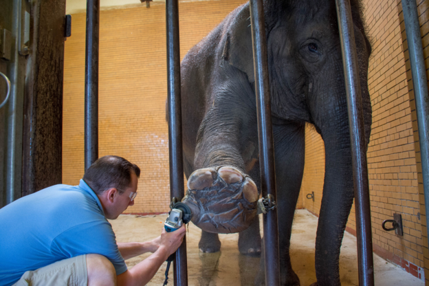
Article
Elephant Foot Health: Detecting Problems Before Lameness
Foot problems in elephants do not necessarily begin with obvious lameness. By the time an elephant i...
Article
When an Elephant’s Behaviour Changes: Recognising Clinically Relevant Patterns
A change in an elephant’s behaviour can be easy to dismiss as temperament. It can also...
Article
Is It Normal for This Elephant? Building an Individual Health Baseline
For a long-lived animal, a single health assessment provides only a snapshot. A more useful c...
Article
Body Condition in Asian Elephants: When Does Change Become Clinically Relevant?
Body condition is often recorded during routine elephant health assessments. Its clinical value, how...
Article
EEHV in Asian Elephant Calves: Recognising the Early Clinical Picture
Elephant endotheliotropic herpesvirus (EEHV) is an important cause of fatal haemorrhagic disease in...
Article
Gastrointestinal Parasites in Asian Elephants: Interpreting Faecal Findings
A positive faecal examination confirms the detection of parasite stages in the sample. It does not,...
Article
Tuberculosis in Asian Elephants: Interpreting Screening Results in Context
Tuberculosis surveillance in elephants presents a fundamental diagnostic challenge: a test result is...
Article
Pregnancy Monitoring in Asian Elephants: Interpreting Ultrasound and Hormonal Findings
Confirming pregnancy is only the beginning of reproductive monitoring in an elephant. With a...