Article
Could Navicular Bone Shape Predict Future Lameness?
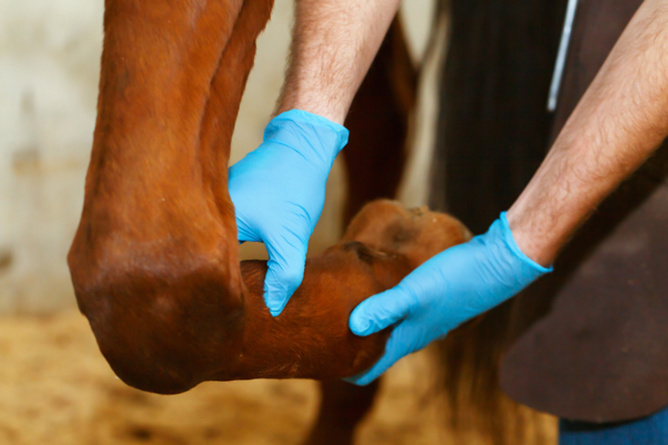
Navicular disease remains one of the most common causes of performance-limiting forelimb lameness in horses, particularly in gallopers, jumpers, Western performance horses, and Quarter Horses. While traditionally viewed as an overuse injury, growing evidence suggests that biomechanics and hereditary bone morphology may play equally important roles in disease development1.
More Than Just a Sesamoid Bone
Although commonly classified as a sesamoid bone, the navicular bone is unique. Unlike typical sesamoid bones embedded within tendons, the navicular bone articulates with both the middle and distal phalanges and functions as a biomechanical pulley for the deep digital flexor tendon (DDFT)2. By maintaining a constant insertion angle of the DDFT, it preserves the tendon’s mechanical advantage and helps absorb concussion forces during locomotion1.
Every stride places substantial compressive forces on the navicular bone. The DDFT wraps around the bone and exerts pressure directly onto its flexor surface, making the navicular apparatus particularly vulnerable to repetitive mechanical stress1.
When Shape Influences Stress
One of the most intriguing findings in recent years is that the shape of the navicular bone may influence how forces are distributed across the foot3.
Research has demonstrated that specific morphological characteristics—including navicular bone position, articular surface angles, and overall bone shape—directly affect joint forces, pressure distribution, and the location of the center of pressure (COP) within the hoof. Horses with unfavorable navicular morphology tend to experience higher peak pressures and more uneven stress distribution across the navicular bone1.
This matters because concentrated loading at the distal aspect of the navicular bone may create focal stress points capable of damaging cartilage and subchondral bone over time. Excessive distal loading has also been linked to pathological changes commonly observed in navicular disease, including alterations in vascular supply, abnormal fluid accumulation within the medullary cavity, and remodeling of nutrient foramina1.
The Genetics Behind the Problem
Not all navicular bones are created equal.
Studies have identified distinct navicular bone shapes—including square, wedge-shaped, and trapezoid configurations—that appear to be inherited traits. Certain bone conformations have also been associated with more severe radiographic changes, particularly those exhibiting a convex proximal articular margin1.
These findings may help explain why navicular disease often shows familial patterns and why some horses appear predisposed despite similar training loads and management practices1.
Why This Matters During Pre-Purchase Exams
Radiographic assessment of the navicular bone has long been a cornerstone of pre-purchase examinations4. However, evaluating morphology may provide information beyond the identification of existing lesions.
Assessment of navicular shape and associated biomechanical parameters could help identify horses that are predisposed to uneven loading patterns before clinical disease develops. This information may be valuable not only for buyers but also for breeders seeking to reduce the transmission of potentially unfavorable conformational traits1.
A Biomechanical Explanation for Navicular Disease?
Previous work demonstrated that horses with navicular disease experience substantially greater compressive forces from the DDFT than sound horses, particularly during the early stance phase. Increased loading appears to be linked to a more cranial center of pressure within the hoof, which lengthens the ground reaction force moment arm and amplifies stress on the navicular apparatus1,5.
Importantly, this suggests that navicular disease may not simply be the primary cause of heel pain. Instead, it may represent the final outcome of chronic abnormal loading within the foot, with navicular bone morphology influencing how well the structure tolerates these forces (31).
Conclusion
Navicular disease is increasingly recognized as a complex interaction between biomechanics, workload, and inherited conformation. While excessive loading remains an important factor, the shape of the navicular bone itself may determine how those forces are distributed. As diagnostic imaging and biomechanical analysis continue to evolve, evaluating navicular morphology may become an important tool for identifying horses at risk long before clinical lameness develops.
References
- Fuss FK. Joint stress analysis of the navicular bone of the horse and its implications for navicular disease. Bioengineering. 2024 Jan 17;11(1):87. https://doi.org/10.3390/bioengineering11010087
- Van Wulfen KK, Bowker RM. Microanatomic characteristics of the insertion of the distal sesamoidean impar ligament and deep digital flexor tendon on the distal phalanx in healthy feet obtained from horses. American journal of veterinary research. 2002 Feb 1;63(2):215-21. https://www.academia.edu/download/90438035/ajvr.2002.63.21520220830-1-1gdfb23.pdf
- Wilson AM, McGuigan MP, Fouracre L, MacMahon L. The force and contact stress on the navicular bone during trot locomotion in sound horses and horses with navicular disease. Equine veterinary journal. 2001 Mar;33(2):159-65. https://www.innovision-systems.com/PDFs/Papers/The%20force%20and%20contact%20stress%20on%20the%20navicular%20bone%20during%20trot%20locomotion.pdf
- Hinkle F, Barrett M. CPD article: Radiographic interpretation of the navicular bone: A review. UK-Vet Equine. 2020 Sep 2;4(5):136-43. https://www.magonlinelibrary.com/doi/abs/10.12968/ukve.2020.4.5.136
- Wilson AM, McGuigan MP, Fouracre L, MacMahon L. The force and contact stress on the navicular bone during trot locomotion in sound horses and horses with navicular disease. Equine veterinary journal. 2001 Mar;33(2):159-65. https://www.innovision-systems.com/PDFs/Papers/The%20force%20and%20contact%20stress%20on%20the%20navicular%20bone%20during%20trot%20locomotion.pdf
Related Contents
Article
Recognising Leptospirosis Across Animal Species: Clinical Presentations and Organ-Specific Changes
A dog presents with fever and systemic illness, while cattle experience reproductive failure without...
Article
Psychogenic Polydipsia in Dogs: Recognising Behavioural Triggers and Clinical Indicators
A dog presents with persistent excessive drinking and urination, yet its physical examination reveal...
Article
Diagnostic Approach to Canine PU/PD: Differentiating Primary Polydipsia from Primary Polyuria
Persistent excessive drinking and urination can be the presenting signs of several conditions in dog...
.jpg)
Upcoming Event
Culture-Guided Treatment of UTIs in Dogs and Cats
Make informed antibiotic choices in canine and feline UTIs with a culture-guided approach. Explore h...
Article
Managing Psychogenic Polydipsia in Dogs: Water Intake, Behavioural Therapy and Pharmacological Intervention
Once psychogenic polydipsia (PPD) has been identified, management needs to address more than the vol...
Article
Duck Viral Hepatitis in Ducklings: Clinical Recognition and Differential Diagnosis
A sudden rise in mortality among young ducklings, particularly those under three weeks of age, shoul...
Article
Confirming Duck Viral Hepatitis: Choosing and Interpreting Diagnostic Tests
When young ducklings develop sudden mortality and liver lesions suggestive of duck viral hepatitis (...
Article
Duck Viral Hepatitis Vaccination: Managing Maternal Antibody Interference and Field Efficacy
Vaccination is a key strategy for controlling Duck Viral Hepatitis (DVH), particularly in intensive...