Article
Tusk Fractures in Asian Elephants: When Pulp Involvement Changes the Clinical Approach
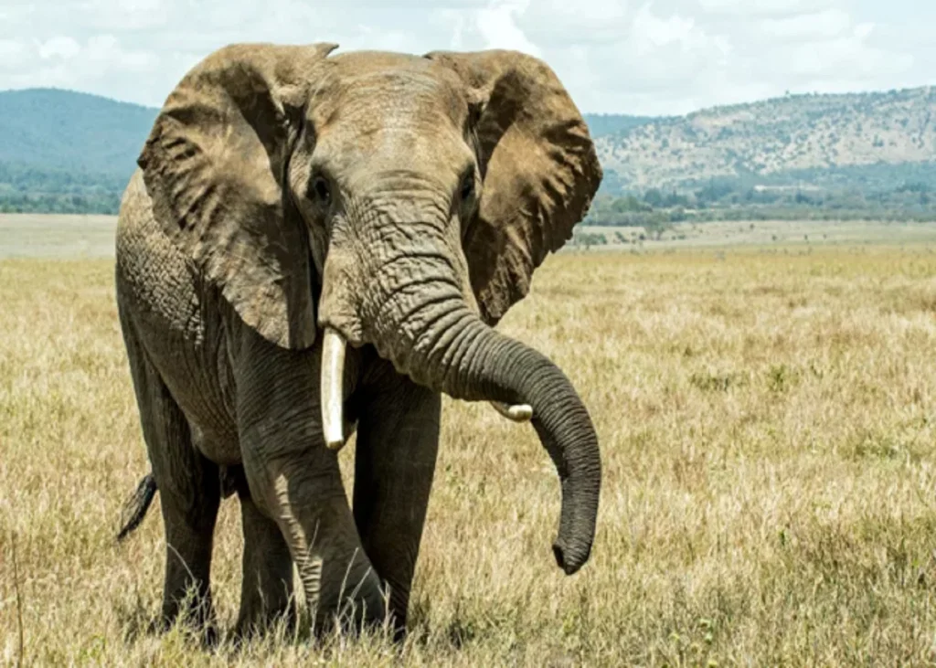
A tusk fracture should not be assessed solely by how much of the tusk has broken.
The more important clinical question is often whether the fracture has exposed or affected the pulp.
This distinction matters because pulp involvement can increase the risk of subsequent pulp disease and may alter decisions regarding treatment, monitoring and tusk preservation.
Begin by defining the fracture
The initial assessment should establish:
- The location and configuration of the fracture;
- Whether the pulp canal is exposed;
- The approximate size of the pulp exposure;
- The condition of the exposed tissue;
- The presence of discharge or swelling;
- Evidence of pulp inflammation or infection;
- The stability of the remaining tusk.
This structured assessment is important because tusk fractures occur under different circumstances and are not a uniform clinical entity.
An international study of managed elephants identified conspecific interactions, tusks becoming caught in enclosure or enrichment structures and impact against objects among the reported causes of traumatic tusk fractures[1].
The study also found that management changes following an initial fracture were associated with a lower likelihood of a subsequent fracture[1].
This gives the clinical history an important role in the assessment. Understanding how the injury occurred may help identify preventable risks alongside addressing the existing fracture.
Pulp exposure changes the level of concern
In a multicentre study of 79 tusk fractures in 64 managed elephants, 44.3% of the fractures involved the pulp[2].
The study included Asian and African elephants. Therefore, its findings provide relevant clinical evidence for Asian elephants but should not be interpreted as representing an exclusively Asian elephant population[2].
Among fractures with pulp involvement, pulp-canal exposures greater than 0.5 cm were associated with a substantially higher likelihood of pulpitis than smaller exposures[2].
This does not mean that exposure size alone determines whether pulpitis will develop.
However, it reinforces the importance of documenting the extent of pulp exposure and monitoring these fractures carefully[2].
Treatment should be guided by the fracture, not by a fixed protocol
A pulp-exposed fracture requires a different level of clinical consideration from a fracture in which the pulp remains protected.
The multicentre study found that, among tusks with pulp involvement, endodontic treatment was associated with a greater likelihood of healing and a reduced likelihood of extraction compared with medical management alone[2].
However, this was an observational study.
The findings should therefore be interpreted as an association rather than evidence that endodontic treatment is required or appropriate for every pulp-exposed tusk[2].
The treatment decision should consider:
- The extent of pulp involvement;
- The condition of the exposed pulp;
- Evidence of pulpitis;
- Fracture stability;
- Contamination;
- The structural integrity of the remaining tusk;
- The elephant's overall clinical status;
- The feasibility of treatment and follow-up.
Conservative management should not be dismissed
The reported outcomes of elephant tusk fractures range from healing with conservative management to the development of serious complications[2].
Therefore, the presence of a fracture alone does not determine the treatment approach.
The clinical objective is to identify which fractures can be monitored and which require more active intervention based on the characteristics and progression of the individual injury.
Follow-up is part of the clinical decision
A fracture that appears stable during the initial examination should not automatically be considered resolved.
Changes associated with pulp disease may develop after the original injury, making continued assessment clinically important[2].
Where feasible, follow-up may include repeat clinical examination, photographic documentation and imaging to evaluate the affected tusk and identify changes over time.
The value of serial assessment lies in comparing the fracture with its previous documented state rather than relying solely on the initial appearance.
Do not overlook prevention after the injury
The fracture itself should be treated as a clinical event, but the circumstances surrounding it may also require investigation.
The epidemiological study identified associations between subsequent fractures and the absence of management changes following the initial injury[1].
This does not establish that a specific management change will prevent every future fracture.
However, it supports reviewing potential risks such as social interactions, enclosure structures and environmental objects after a fracture has occurred[1].
Clinical takeaway
The presence or absence of pulp involvement is a key factor in the clinical assessment of a fractured tusk.
Pulp-exposed fractures require careful evaluation and structured follow-up because of the potential for subsequent pulp disease.
Current evidence also suggests that endodontic treatment may improve outcomes in selected fractures with pulp involvement, but the available data support individualised clinical decision-making rather than a single treatment protocol for every tusk fracture.
References:
- Rose JB, Leeds A, Yang LM, et al. Treatment and Outcomes of Tusk Fractures in Managed African Savanna and Asian Elephants (Loxodonta africana and Elephas maximus) across Five Continents. Animals (Basel). 2022;12(9):1125. Published 2022 Apr 27. https://doi.org/10.3390/ani12091125
- Rose JB, Leeds A, Yang LM, LeMont R, Fayette MA, Proudfoot JS, Bowman MR, Woody A, Oosterhuis J, Fagan DA. Treatment and outcomes of tusk fractures in managed African savanna and Asian elephants (Loxodonta africana and Elephas maximus) across five continents. Animals. 2022 Apr 27;12(9):1125. https://doi.org/10.3390/ani12091125
Related Contents
Article
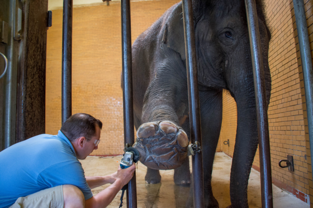
Elephant Foot Health: Detecting Problems Before Lameness
Foot problems in elephants do not necessarily begin with obvious lameness. By the time an elephant i...
Article
When an Elephant’s Behaviour Changes: Recognising Clinically Relevant Patterns
A change in an elephant’s behaviour can be easy to dismiss as temperament. It can also...
Article
Is It Normal for This Elephant? Building an Individual Health Baseline
For a long-lived animal, a single health assessment provides only a snapshot. A more useful c...
Article
Body Condition in Asian Elephants: When Does Change Become Clinically Relevant?
Body condition is often recorded during routine elephant health assessments. Its clinical value, how...
Article
EEHV in Asian Elephant Calves: Recognising the Early Clinical Picture
Elephant endotheliotropic herpesvirus (EEHV) is an important cause of fatal haemorrhagic disease in...
Article
Gastrointestinal Parasites in Asian Elephants: Interpreting Faecal Findings
A positive faecal examination confirms the detection of parasite stages in the sample. It does not,...
Article
Tuberculosis in Asian Elephants: Interpreting Screening Results in Context
Tuberculosis surveillance in elephants presents a fundamental diagnostic challenge: a test result is...
Article
Pregnancy Monitoring in Asian Elephants: Interpreting Ultrasound and Hormonal Findings
Confirming pregnancy is only the beginning of reproductive monitoring in an elephant. With a...