Article
Contagious Ecthyma in Sheep and Goats: Recognizing Lesions and Differentiating Orf in Practice
Contagious ecthyma, also known as orf, scabby mouth, sore mouth, or contagious pustular dermatitis, is an acute, contagious skin disease affecting sheep, goats, and several other ruminants. The disease is caused by Orf virus (ORFV), a member of the genus Parapoxvirus within the family Poxviridae. Although mortality is generally low, morbidity can be substantial, particularly when painful lesions interfere with feeding and nursing1.
For the practicing veterinarian, recognition of the characteristic proliferative lesions and their progression is an important first step in distinguishing contagious ecthyma from other diseases producing oral, facial, or generalized skin lesions.
Recognizing the Clinical Pattern
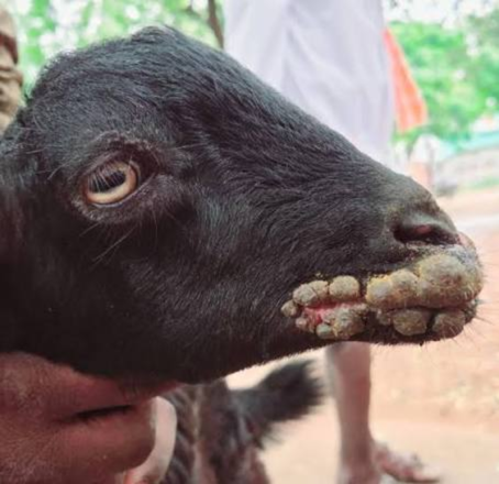
Lesions commonly develop after an incubation period of several days. The PDF describes an incubation period ranging from 4–8 days, with an initial increase in body temperature followed by papules and pustules, particularly around the lips, nose, and oral commissures. Lesions may subsequently become thick, sticky, and inflammatory.
Earlier descriptions in the same source report an incubation period of 8–14 days, with proliferative lesions appearing on the lips, snout, ears, eyelids, tongue, and nostrils. Lesions can occasionally extend to non-woolly areas such as the legs, feet, and udder. Their progression is described from redness and blisters through bumps and crusts to sores that gradually improve over one to two months2.
This progression is clinically important because the appearance of the lesion can vary with its stage. Proliferative, persistent nodular lesions are considered characteristic of orf and may occur in labial, generalized, mammary, or vaginal forms.
Clinical Impact on Lambs and Kids1
The location of lesions can have a direct effect on animal performance. Lesions involving the mouth are painful, and affected lambs and young animals may experience difficulty grazing or nursing. The source notes that lesions can result in anorexia followed by starvation, while thin scabs may be susceptible to bleeding.
Orf is not always restricted to superficial skin changes. Secondary bacterial involvement may complicate the clinical picture. Dermatophilus congolensis can make accurate diagnosis more difficult, while buccal mucosal lesions associated with Fusobacterium necrophorum may disseminate to visceral sites. Such involvement has been associated with granulomatous lesions, hoof shedding, and vulval ulcers in generalized disease.
Histopathological Features
The histological appearance varies according to the developmental stage of the lesion. Skin and mucosal lesions may present as widespread vesicular-papular changes. The source describes prominent rete ridges, substantial parakeratotic and orthokeratotic hyperkeratosis, and considerable epidermal hyperplasia as characteristic histological alterations. Lesions rarely exceed 5 cm in diameter, although lesions progressing through different clinical stages may measure greater than 1–3 cm3.
Differential Diagnosis: Look Beyond the Lesion1
The clinical appearance of contagious ecthyma can overlap with other important diseases. Differential diagnosis should include ulcerative dermatosis, sheep pox, and foot-and-mouth disease (FMD).
A useful clinical distinction from FMD is the proliferative nature of orf lesions. In sheep affected by FMD, oral lesions are more likely to involve the lower gums and tongue, whereas proliferative lesions are a prominent feature of orf. Ulcerative dermatosis may produce inflammation and crust formation involving the face, feet, and genitalia, while sheep and goat pox are characterized by elevated papules distributed throughout the body.
Practical Clinical Insight
When confronted with facial or oral lesions in sheep or goats, consider lesion character, distribution, progression, age of affected animals, and interference with feeding or nursing together rather than relying on a single visible lesion. The characteristic proliferative pattern supports clinical recognition of contagious ecthyma, but overlapping presentations and secondary bacterial involvement mean that laboratory confirmation may be required for definitive validation.
References
- Bhagawati N, Shankarishan P, Habibi M. Contagious ecthyma in goat and sheep: a review of current status and future perspectives. Uttar Pradesh J Zool. 2025;46(10):229-239. doi:10.56557/UPJOZ/2025/v46i104982. https://mbimph.com/index.php/UPJOZ/article/view/4982/6394
- Peralta A, Robles CA, Micheluod JF, Rossanigo CE, Martinez A, Carosio A, König GA. Phylogenetic analysis of ORF viruses from five contagious ecthyma outbreaks in Argentinian goats. Frontiers in veterinary science. 2018 Jun 19;5:134. https://www.frontiersin.org/journals/veterinary-science/articles/10.3389/fvets.2018.00134/pdf
- McElroy MC, Bassett HF. The development of oral lesions in lambs naturally infected with orf virus. The Veterinary Journal. 2007 Nov 1;174(3):663-4. https://doi.org/10.1016/j.tvjl.2006.10.024
Related Contents
.jpg)
Video
Transjugular Removal of Canine Heartworms: A Clinical and Surgical Overview
Transjugular removal of canine heartworms is a specialized intervention used in selected dogs with s...

Article
Feline Pregnancy Endocrinology: Hormonal Changes and Placental Function
Pregnancy in cats involves a distinctive endocrine pattern in which ovarian and placental hormones w...

Article
Hormonal Control of Pregnancy Termination in Cats
Pregnancy maintenance in cats depends strongly on coordinated hormonal support, particularly progest...

Article
Feline Embryo Implantation and Early Placental Development
Pregnancy establishment in cats involves a sequence of precisely timed events between fertilization,...

Article
Ultrasonographic Monitoring of Pregnancy in Cats: A Gestational Timeline
Ultrasonography provides a practical way to follow the progression of feline pregnancy by identifyin...

Article
Feline Mismating: Pregnancy Diagnosis and Clinical Management
Mismating or unplanned mating is a common clinical problem in sexually mature queen cats because of...

Article
Medical Termination of Pregnancy in Queens: Drugs, Timing and Treatment Protocols
Medical termination of pregnancy (MTP) in queen cats should be approached according to the timing of...

Article
Safety and Aftercare Following Pregnancy Termination in Queen Cats
Pregnancy termination in queen cats requires more than selecting an appropriate abortifacient. Once...