Article
Parasitic Diseases in Farmed Fish: Recognising Gill, Skin and Systemic Involvement
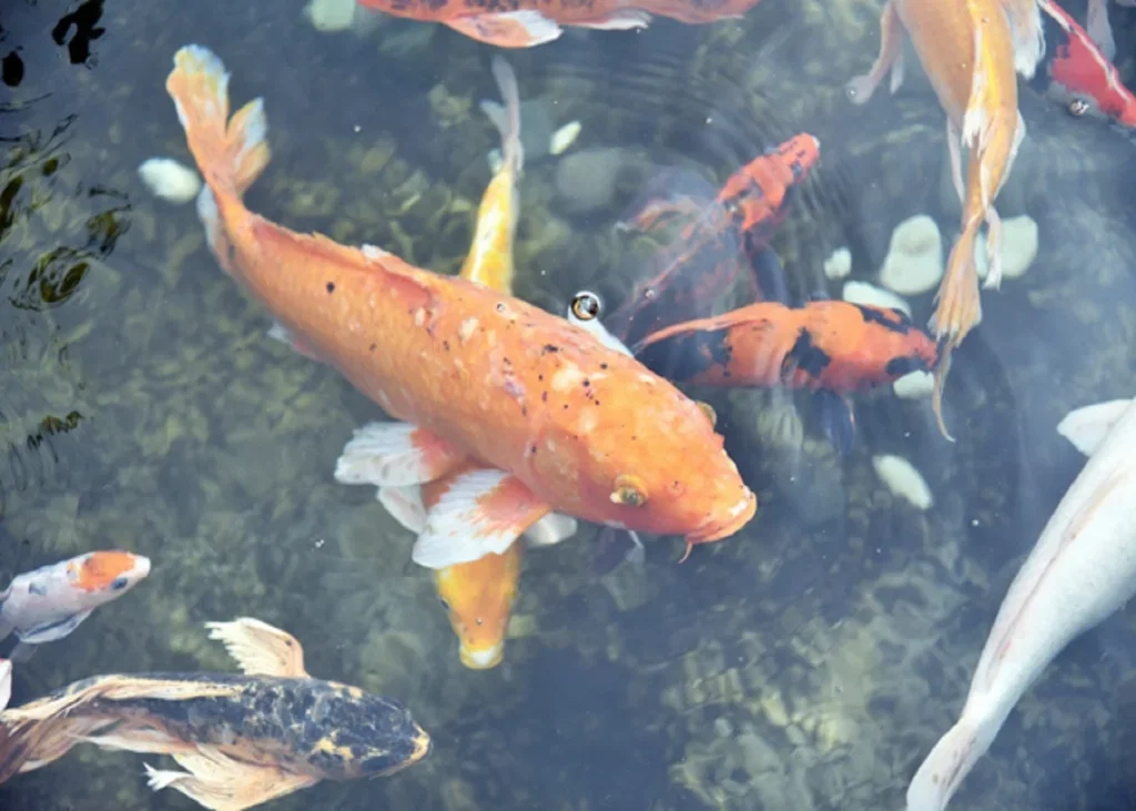
A group of fingerlings is losing appetite and becoming increasingly inactive. Some fish have excessive mucus, while others show darkened skin or visible white spots.
When several external signs appear together, where should the clinical assessment begin?
Parasitic infections are important causes of disease in cultured fish and can interfere with growth and overall health. The clinical presentation varies with the parasite involved and the tissues affected. For practicing veterinarians, recognising whether the gills, skin or other tissues are primarily involved provides an important starting point for assessment.
Start with the affected tissue
Parasites injure their hosts while deriving substances from them. Those with direct life cycles are important pathogens of farmed fish, while parasites with indirect life cycles use fish as an intermediate host[1].
The location of the parasite can strongly influence clinical presentation. Gill involvement may compromise respiratory function, while skin infestation can produce excessive mucus, colour changes or visible lesions.
This makes the distribution of clinical signs particularly useful during examination.
White spots: Look beyond the skin
Ichthyophthirius multifiliis, responsible for ichthyophthiriasis or white spot disease, is an important protozoan parasite of freshwater fish, particularly Indian major carp fry and fingerlings. It can affect fish at different stages, from fry to brood fish, and is associated with losses in ornamental and freshwater cultured fish[2].
The parasite affects epithelial tissues of the gills, fins and skin, producing the characteristic visible white spots.
But the clinical impact is not limited to what can be seen externally. Gill and epithelial damage can interfere with respiratory, osmoregulatory and excretory functions.
Affected fish may become:
- Lethargic
- Clamped-finned
- Dark in colour
Therefore, visible white spots should prompt assessment for the broader effects of the infestation rather than being treated simply as a superficial skin finding.
When the skin looks grey or blue
A different external pattern is associated with Ichthyobodo necator, the causative agent of ichthyobodosis.
This ectoparasitic flagellate affects both freshwater and marine fish, including ornamental fish, and particularly affects the gills and skin of salmonid fry and fingerlings[3].
The skin may develop a steel-grey appearance, accompanied by grey or blue-coloured mucus. Weakness, inactivity and loss of appetite may also occur.
The combination is clinically useful: A grey or blue mucus-covered fish with weakness and poor appetite presents a different pattern from the characteristic white spots associated with I. multifiliis.
Excess mucus and gill involvement
Trichodina spp. are ciliated ectoparasites found in both freshwater and marine fish. They are among the common ectoparasites affecting cultivated and wild fish and involve the skin and gills[2].
Affected fish may develop respiratory distress, loss of appetite and scales, along with black skin. Excessive mucus accumulation in the gill pouches can also occur.
Here, the gills deserve particular attention. Respiratory distress combined with excessive mucus can indicate that the parasitic burden is affecting more than the external appearance of the fish.
Clinical bottom line
Parasitic disease should not be assessed solely by looking for a named parasite or a single characteristic lesion.
Instead, ask:
Where is the clinical problem concentrated—skin, gills or both?
White spots with lethargy and clamped fins suggest a different clinical pattern from steel-grey skin with blue-grey mucus, while respiratory distress with excessive mucus can point towards another ectoparasitic presentation.
For routine fish-health assessment, combining affected tissue, colour changes, mucus production, appetite and behaviour can provide a practical framework for recognising parasitic disease and narrowing the clinical differential.
References:
- Faruk MA. Fish parasite: infectious diseases associated with fish parasite. InSeafood Safety and Quality 2018 Aug 6 (pp. 154-176). CRC Press. https://www.researchgate.net/profile/Md-Faruk-5/publication/326345217
- Senthamarai MD, Rajan MR, Bharathi PV. Current risks of microbial infections in fish and their prevention methods: A review. Microbial pathogenesis. 2023 Dec 1;185:106400. https://www.researchgate.net/profile/Rajan-Muthuswami-Ruby/publication/375006029
- Isaksen TE. Ichthyobodo infections on farmed and wild fish. Bergen: University of Bergen. 2013. https://www.researchgate.net/profile/Trond-Isaksen-2/publication/333402520
Related Contents

Article
Chronic Cough in Dogs and Cats: How to Classify and Localize It
A dog has been coughing for several weeks. The owner describes it as “honking,” but the...

Article
Diagnosing the Coughing Dog or Cat: Choosing the Right Diagnostic Test
A coughing dog or cat does not automatically need every available respiratory test. The more useful...

Article
Bronchoscopy and Airway Sampling in Dogs and Cats with Chronic Cough
In dogs and cats with chronic cough, thoracic radiographs may not fully explain the airway abnormali...

Article
When Should Veterinarians Suppress Cough?
Cough suppression plays a specific role in managing respiratory diseases in dogs and cats. The aim i...

Article
Managing Persistent Cough in Dogs and Cats: From Conventional Therapy to Emerging Options
Persistent cough in dogs and cats may require more than treatment of the primary disease. When cough...
Article
Bacterial Diseases in Farmed Fish: Recognising Clinical Patterns in Practice
A fish population develops haemorrhagic lesions, lethargy and poor appetite. Another shows exophthal...
Article
Viral Diseases in Aquaculture: Recognising Clinical Patterns Before the Outbreak Escalates
A group of cultured fish begins showing dark skin, haemorrhage, anaemia and abnormal behaviour. In a...
Article
Fungal and Oomycete Infections in Fish: Recognising Environmental and Clinical Triggers
Fungal disease in a fish population may appear after another illness, mechanical injury, or a period...