Article
When a Mild Uterine Torsion Turns Dangerous: Lessons from a Late-Gestation Mare
Not every uterine torsion in a mare presents as a dramatic emergency. Some appear mild, resolve spontaneously, and initially seem clinically stable. But as this case highlights, even a low-grade torsion can evolve into a life-threatening reproductive disaster if subtle warning signs are missed1.
Uterine torsion accounts for approximately 5–10% of severe dystocia cases in mares, with many occurring before term gestation1. Clinical signs can range from mild abdominal discomfort to severe colic, depending on the degree of vascular compromise and uterine rotation2.
The Initial Presentation
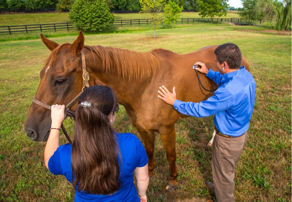
An 11-year-old primiparous Arabian mare at 331 days of gestation was examined for abdominal pain. Interestingly, the owner also reported an earlier episode of colic around day 296 that had been resolved after NSAID administration1.
Clinical examination showed:
- Heart rate: 60 bpm
- Respiratory rate: 22 breaths/min
- Normal rectal temperature
- No vaginal discharge
- Closed cervix
Transrectal palpation revealed a clockwise uterine torsion of approximately 90°, identified through displacement and tension of the broad ligaments. Ultrasonography confirmed that the fetus was still viable.
Because the torsion was relatively mild, medical management with flunixin meglumine was initiated.
When “Improvement” Can Be Misleading
The following day, the uterus had returned to a normal anatomical position spontaneously. Repeat ultrasonography showed an active fetus with normal movement patterns1.
Five days later, fetal viability was still confirmed.
At this stage, everything appeared encouraging:
- The torsion had corrected itself
- The mare was clinically stable
- No systemic compromise was evident
- Fetal activity remained normal
But this is where the case becomes clinically important.
Spontaneous correction of uterine torsion does not always mean the uterus escaped injury.
Why Monitoring Matters
As gestation progressed, the mare failed to foal by day 350. The owner also noticed involution of the mammary glands — an abnormal finding in a near-term mare.
Repeat ultrasonography revealed a shocking development:
- The uterus was empty and involuted
- The fetus was dead
- The fetus was floating freely within the abdominal cavity
The mare had developed secondary abdominal pregnancy following uterine rupture1.
The Hidden Risk in Late-Gestation Torsion
The exact mechanism of rupture was unclear, but clinicians suspected that torsion may have partially compromised uterine blood flow, resulting in fetal distress and excessive fetal movement.
Mechanical trauma from vigorous fetal forelimb activity may have contributed to tearing of the uterine wall.
This case reinforces an important clinical reality:
Even apparently mild uterine torsions can progress to catastrophic complications later in gestation1.
The Role of Ultrasonography
One of the strongest lessons from this case was the importance of advanced ultrasonographic monitoring.
Late-gestation transabdominal ultrasonography in mares is technically demanding. Evaluating:
- uterine integrity,
- fetal positioning,
- fetal viability,
- abdominal fluid,
- and displaced fetal structures
requires significant operator experience.
Because the mare initially appeared stable, the severity of the complication was not immediately recognized.
The authors emphasized that more intensive serial ultrasonographic evaluations — ideally during hospitalization — may have allowed earlier detection of progressive abnormalities1.
Practical Take-Home Messages for Veterinarians
Never underestimate mild torsion
A 90° torsion may appear clinically manageable but can still damage the uterus.
Repeated imaging is critical
Single examinations may miss evolving pathology.
Watch for subtle late-gestation changes
Failure to foal, mammary involution, or reduced fetal activity should trigger immediate reassessment.
Hospitalization improves monitoring
Continuous observation may identify deterioration earlier than field management.
Experience matters
Late-term reproductive ultrasonography requires advanced interpretation skills.
Conclusion
This case demonstrates how a seemingly uncomplicated uterine torsion can silently progress into a severe reproductive emergency. The absence of dramatic clinical signs delayed recognition of uterine rupture and abdominal pregnancy.
For equine practitioners, the message is clear: mares with late-gestation uterine torsion deserve aggressive monitoring even after apparent clinical improvement.
References
- Palmieri V, Catone G, Conte A, Di Palma T, Liguori P, Izzo I, Vullo C. Near-Term Extrauterine Pregnancy Secondary To Uterine Rupture In A Mare. Journal of Equine Veterinary Science. 2026 May 22:105943. https://www.sciencedirect.com/science/article/pii/S0737080626001784
- MURUGAN, M., KUMAR, K.S., NAPOLEAN, R.E., GOPIKRISHNAN, D., SELVARAJU, M., RAVIKUMAR, K., PALANISAMY, M. and DHARMACEELAN, S., AN UNUSUAL CASE OF UTERINE TORSION AND ITS MANAGEMENT BY LAPAROHYSTEROTOMY IN A KATHIAWARI MARE. Haryana Veterinarian, 62(2), pp.148-149. https://www.luvas.edu.in/haryana-veterinarian/download/dec-2023/32.pdf
Related Contents
Article
Recognising Leptospirosis Across Animal Species: Clinical Presentations and Organ-Specific Changes
A dog presents with fever and systemic illness, while cattle experience reproductive failure without...

Article
Psychogenic Polydipsia in Dogs: Recognising Behavioural Triggers and Clinical Indicators
A dog presents with persistent excessive drinking and urination, yet its physical examination reveal...

Article
Diagnostic Approach to Canine PU/PD: Differentiating Primary Polydipsia from Primary Polyuria
Persistent excessive drinking and urination can be the presenting signs of several conditions in dog...
.jpg)
Upcoming Event
Culture-Guided Treatment of UTIs in Dogs and Cats
Make informed antibiotic choices in canine and feline UTIs with a culture-guided approach. Explore h...

Article
Managing Psychogenic Polydipsia in Dogs: Water Intake, Behavioural Therapy and Pharmacological Intervention
Once psychogenic polydipsia (PPD) has been identified, management needs to address more than the vol...

Article
Duck Viral Hepatitis in Ducklings: Clinical Recognition and Differential Diagnosis
A sudden rise in mortality among young ducklings, particularly those under three weeks of age, shoul...
Article
Confirming Duck Viral Hepatitis: Choosing and Interpreting Diagnostic Tests
When young ducklings develop sudden mortality and liver lesions suggestive of duck viral hepatitis (...
Article
Duck Viral Hepatitis Vaccination: Managing Maternal Antibody Interference and Field Efficacy
Vaccination is a key strategy for controlling Duck Viral Hepatitis (DVH), particularly in intensive...